Nature | Art | Science
Diatoms, coloured scanning electron micrograph (SEM). Coscinodiscus marginatus (left) and Eupyxidicula sp. (right). Shows to the intricate silica-based cell walls of two fossil diatoms, showcasing the beauty and complexity of these microscopic organisms. Diatoms, a major group of algae found in the oceans, waterways, and soils of the world, are not just marvels of natural art; they are critical to our planet's ecology. They contribute significantly to the oxygen in the atmosphere and are a fundamental link in the aquatic food chain. Diatoms are among the most diverse species on earth. Estimates of the number of diatom species range from 20.000-200.000. Scientists are discovering new species every year. Magnification: x600 when printed 10 centimetres wide.
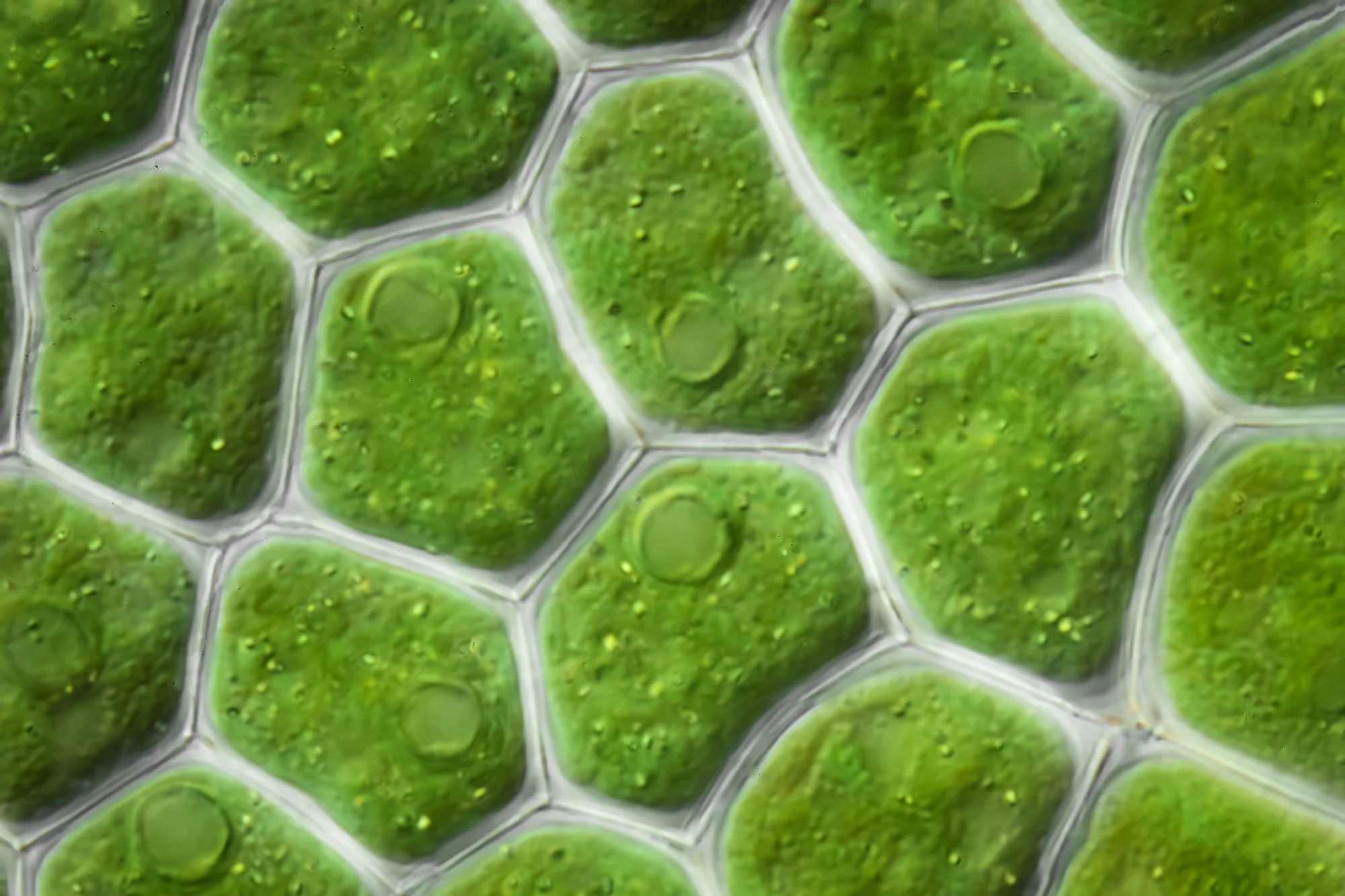
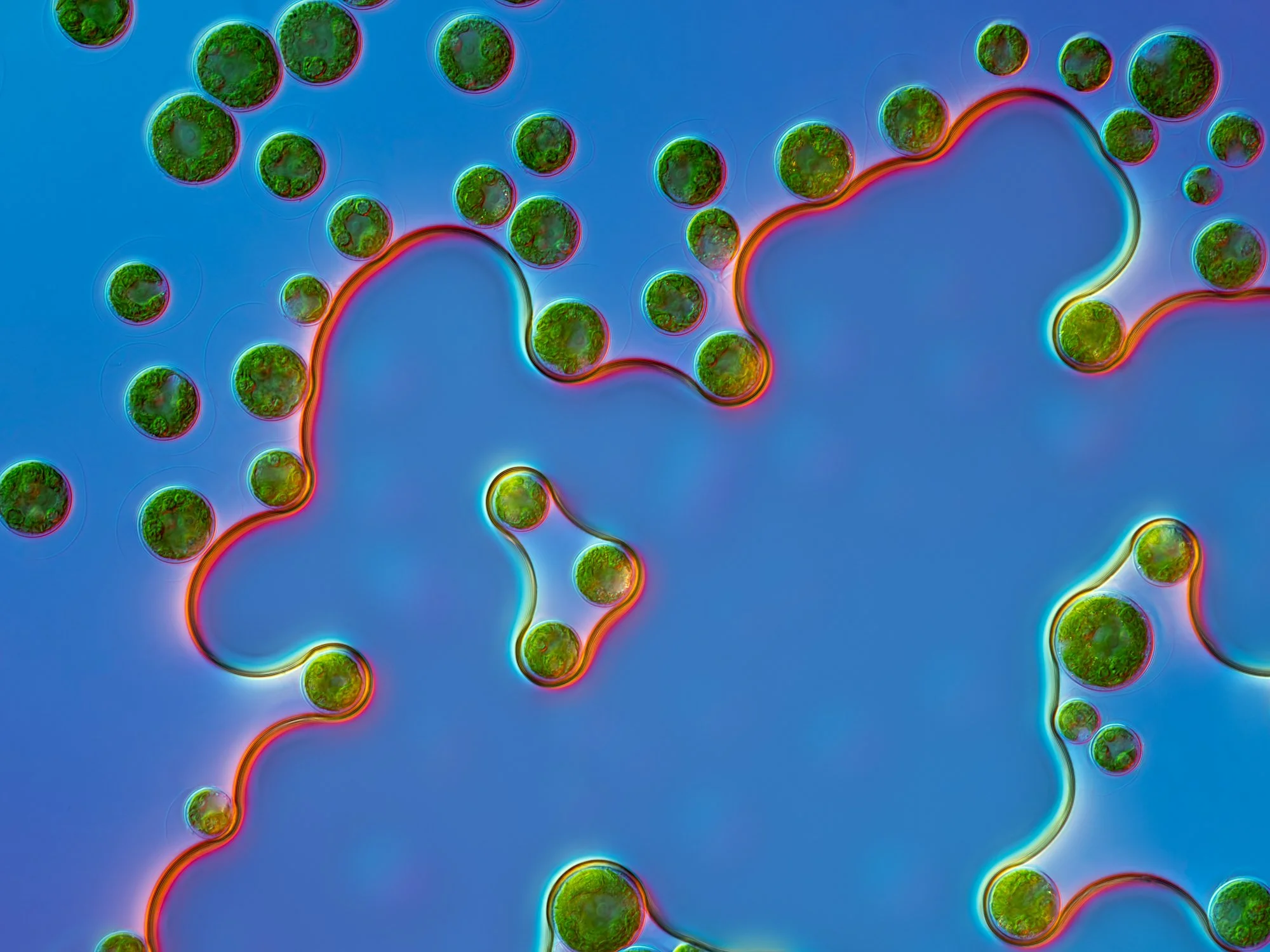
Differential interference contrast (DIC) light micrograph of a green alga Pediastrum duplex. Pediastrum is a very distinctive coenobial colony with a flattened, often starlike shape. The outer half of the marginal cells extendinto two horn-like processes with a tuft of long bristles (as seen in the photograph). Magnification: x475 when printed at 10 centimetres wide at its longest edge.
Differential interference contrast light micrograph of a golden algae Dinobryon sp. Golden algae are a large group of algae, found mostly in freshwater environments. In this colony individual cells, each cell is propelled by two flagella pulling the entire colony through the water in order to find good light. Dinobryon sp. is a mixotrophic organism, meaning that it is photosynthetic as well ashaving the ability to ingest food. When the light energy or nutrients are limited, it starts eating bacteria to survive. Each colony can eat thousands of bacteria every hour, providing as much as 50% of its energy requirements. Dinobryon is a potent force in controlling bacterial numbers in freshwater lakes and ponds. Magnification: x425 when printed at 10 centimetres wide at its longest edge.
Light micrograph (fluorescence microscopy) of Spirogyra sp. filamentous green algae. Spirogyra is named for the spiral arrangement of its chloroplasts. Its cylindrical cells connect end to end in long green filaments. In this photograph fluorescent dyes were used to highlight various features of the algae. The chloroplasts auto-fluoresce in red due to the ultraviolet light used to illuminate the algae. Magnification: x290 when printed at 10 centimetres wide at its longest edge.
Darkfield light micrograph (polarized light) of a Micrasterias sp. desmid green algae. Desmids are a common group of freshwater single-celled algae that have intricate cell walls. Micrasterias are highly adapted to nutrient-poor, acidic water. Magnification: x190 when printed at 10 centimetres wide at its longest edge.
Differential interference contrast (DIC) light micrograph of a green alga Xanthidium antilopaeum (center). The smaller algae in the background are Istmochloron sp. Xanthidium belongs to a group of fresh-water single-celled algae called desmids which that have intricate cell walls. Magnification: x425 when printed at 10 centimetres wide at its longest edge.
Differential interference contrast light micrograph of two genus of green algae. Pandorina sp. (right) and Eudorina sp. (left). They are microscopic green algae living in colonies and swim/rotate using flagella. The red dots are eye spots to allow them to swim towards the light. The colony is surrounded by a translucent mucilage. Magnification: x600 when printed at 10 centimetres wide at its longest edge.
Differential interference contrast (DIC) light micrograph of the colonial green algae “Botryococcus braunii”. Colonies are held together by a lipid (oily) biofilm as can be seen as oil droplets around the edge of the colony. Typically around 30–40% of the dry weight of a colony is oil. Botryococcus braunii has potential for algaculture because of the hydrocarbons it produces, which can be chemically converted into fuels. Magnification: x425 when printed at 10 centimetres wide at its longest edge.
Light micrograph (polarized light) of Cosmarium sp. Cosmarium are photosyntethesizing microscopic algae that inhabit the upper sunlit layer of almost all bodies of freshwater on earth. Cosmarium are purely planktonic and cannot propel themselves. They are wanderers that drift with the currents. Cosmarium, along with other microscopic green algae, serve as the base of the aquatic food web, providing an essential ecological function for all aquatic life. There are more than 400 different species of Cosmarium. Magnification: x800 when printed at 10 centimetres wide at its longest edge.
Differential interference contrast (DIC) light micrograph of a green alga desmid of the species Euastrum verrucosum. Desmids are a common group of freshwater single-celled algae that have intricate cell walls with curved cell lobes. Magnification: x475 when printed at 10 centimetres wide at its longest edge.
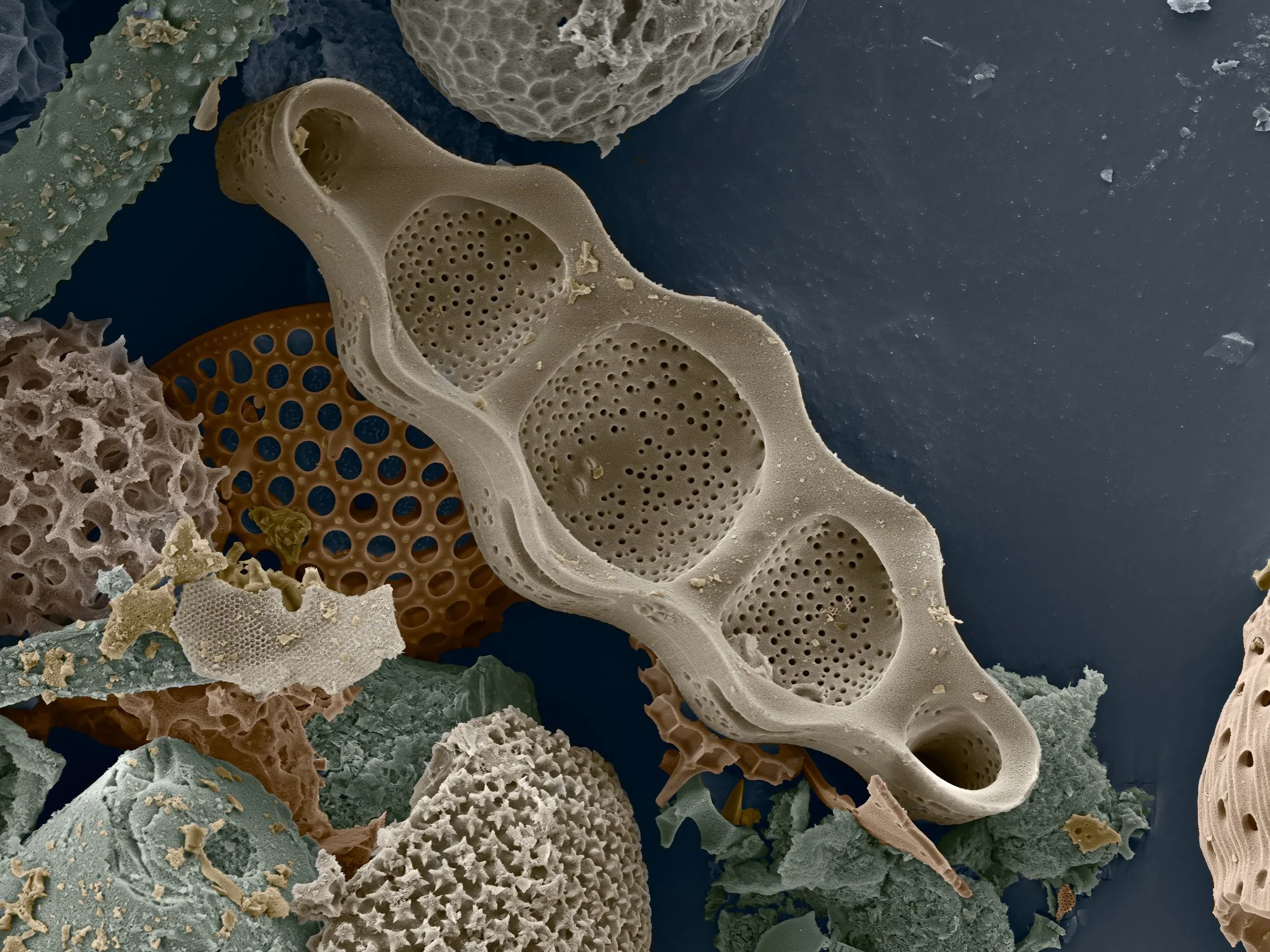
Light micrograph (DIC) of of diatoms of species Tabellaria flocculosa. Diatoms are single-celled photosynthetic algae, of which there are about 100,000 species. They have mineralised cell walls (frustules) that contain silica and provide protection and support. They form an important part of the plankton at the base of the marine and freshwater food chains. Magnification: x450 when printed at 10 centimetres wide at its longest edge.
SEM
Diatom. Coloured scanning electron micrograph (SEM). The photograph shows the genus Gomphonema sp. Diatoms contribute significantly to the oxygen in the atmosphere and are a fundamental link in the aquatic food chain. Diatoms are among the most diverse species on earth. Estimates of the number of diatom species range from 20.000-200.000. Scientists are discovering new species every year. Magnification: x1260 when printed 10 centimetres wide
Diatom. Coloured scanning electron micrograph (SEM). The photograph shows the genus Gomphonema sp. Diatoms contribute significantly to the oxygen in the atmosphere and are a fundamental link in the aquatic food chain. Diatoms are among the most diverse species on earth. Estimates of the number of diatom species range from 20.000-200.000. Scientists are discovering new species every year. Magnification: x1260 when printed 10 centimetres wide
Diatom. Coloured scanning electron micrograph (SEM). Species Didymosphenia geminata, inside valve view. Also colloquially known as 'rock snot' due to its capability to form thick, mucilaginous mats on the bottom of rivers and streams. Characterized by its stalked colonies and distinctive morphology, D. geminata is a subject of ecological concern because of its tendency to bloom excessively under certain environmental conditions. These blooms can cover extensive areas of substrate, impacting aquatic ecosystems by altering habitat structures, nutrient cycling, and water flow. Diatoms contribute significantly to the oxygen in the atmosphere and are a fundamental link in the aquatic food chain. Diatoms are among the most diverse species on earth. Estimates of the number of diatom species range from 20.000-200.000. Scientists are discovering new species every year. Magnification: x680 when printed 10 centimetres wide.
Diatom. Coloured scanning electron micrograph (SEM). Species Didymosphenia geminata, Close-up of outside valve view. Also colloquially known as 'rock snot' due to its capability to form thick, mucilaginous mats on the bottom of rivers and streams. Characterized by its stalked colonies and distinctive morphology, D. geminata is a subject of ecological concern because of its tendency to bloom excessively under certain environmental conditions. These blooms can cover extensive areas of substrate, impacting aquatic ecosystems by altering habitat structures, nutrient cycling, and water flow. Diatoms contribute significantly to the oxygen in the atmosphere and are a fundamental link in the aquatic food chain. Diatoms are among the most diverse species on earth. Estimates of the number of diatom species range from 20.000-200.000. Scientists are discovering new species every year. Magnification: x5500 when printed 10 centimetres wide.
SEM
SEM
SEM
SEM
Differential interference contrast (DIC) light micrograph of a desmid, Closterium sp. The desmids are a group of unicellular green algae characterised by their beautifully shaped cell walls. The cell is split into two halves, joined by a waist. Cell division is accomplished by splitting in two at the waist, with each half generating a perfect replica of itself to restore the original shape. Magnification: x450 when printed at 10 centimetres wide at its longest edge.
Light micrograph (DIC) of of diatoms of species Tabellaria flocculosa. Diatoms are single-celled photosynthetic algae, of which there are about 100,000 species. They have mineralised cell walls (frustules) that contain silica and provide protection and support. They form an important part of the plankton at the base of the marine and freshwater food chains. Magnification: x700 when printed at 10 centimetres wide at its longest edge.
Differential interference contrast (DIC) light micrograph of a Micrasterias sp. desmid green algae. Desmids are a common group of freshwater single-celled algae that have intricate cell walls. Micrasterias are highly adapted to nutrient-poor, acidic water. light micrograph of a Micrasterias sp. desmid green algae. Desmids are a common group of freshwater single-celled algae that have intricate cell walls. Micrasterias are highly adapted to nutrient-poor, acidic water. This cell is undergoing cell division. Magnification: x150 when printed at 10 centimetres wide at its longest edge.
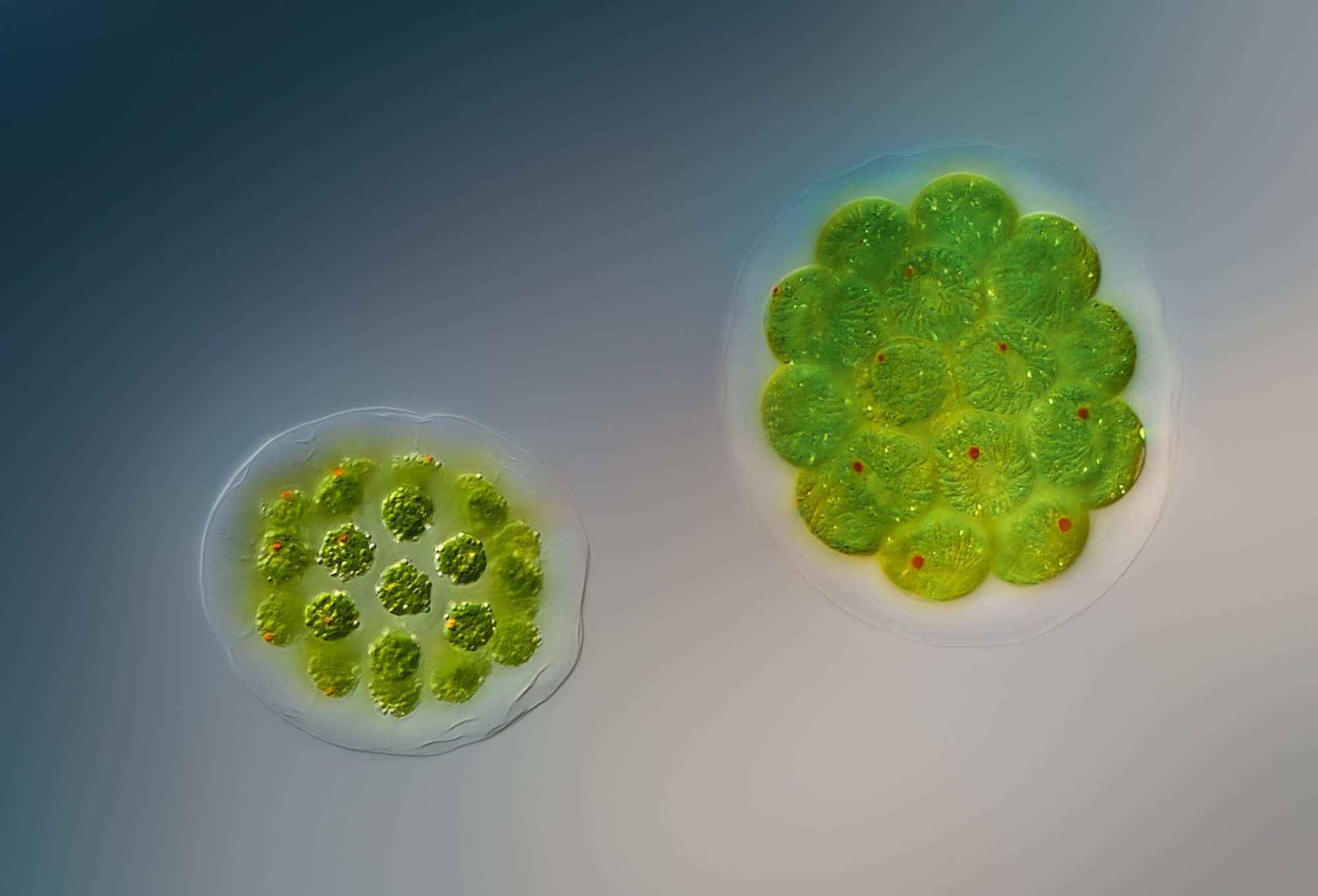
Light micrograph (DIC) of the disc-like colony of cells formed by the green alga, Pediastrum sp. Pediastrum is a type of freshwater colonial algae. The colony comprises a central cell surrounded by concentric rings of cells. Cells (with chloroplast) and cell walls are shown with some cells having visible pyrenoids (circular spots). The pyrenoid's function is to promote photosynthetic CO2 fixation. Magnification: x1400 when printed at 10 centimetres wide at its longest edge.
Light micrograph (DIC) of the disc-like colony of cells formed by the green alga, Pediastrum integrum. Pediastrum is a type of freshwater colonial algae. The colony comprises a central cell surrounded by concentric rings of cells. Cells (with chloroplast) and cell walls are shown with some cells having visible pyrenoids (circular spots). The pyrenoid's function is to promote photosynthetic CO2 fixation. Magnification: x1400 when printed at 10 centimetres wide at its longest edge.
Light micrograph (DIC) of the disc-like colony of cells formed by the green alga, Pediastrum integrum. Pediastrum is a type of freshwater colonial algae. The colony comprises a central cell surrounded by concentric rings of cells. The outermost cells each have two lobes projecting outwards, giving the colony a "spiky" appearance. Magnification: x750 when printed at 10 centimetres wide at its longest edge.
Gigapixel
Gigapixel
Gigapixel
Haematococcus pluvalis. DIC
Haematococcus pluvalis. DIC
Differential interference contrast (DIC) light micrograph of freshwater algae Gonyostomum sp. Gonyostomum can be a nuisance during summers when they bloom. The surface of the Gonyostomum is covered by small spheres of chloroplasts. Inside the cell, it has various vacuoles. Magnification: x500 when printed at 10 centimetres wide at its longest edge.
Differential interference contrast (DIC) light micrograph of a freshwater alga Gonyostomum sp. Gonyostomum can be a nuisance during summers when they bloom. The surface of the Gonyostomum is covered by small spheres of chloroplasts. Inside the cell, it has various vacuoles. The alga uses flagella to propel itself. Magnification: x1250 when printed at 10 centimetres wide at its longest edge.
Light micrograph (fluorescence microscopy) of Spirulina sp. cyanobacteria filaments. Each filament is a colony of bacterial cells. Cyanobacteria are photosynthesizing bacteria that are found in most habitats where water is present. They are also used as a health food and dietary supplement because of their high protein content. Magnification: x400 when printed at 10 centimetres wide at its longest edge.
Polarized light micrograph of a Micrasterias sp. desmid green algae. Desmids are a common group of freshwater single-celled algae that have intricate cell walls. Micrasterias are highly adapted to nutrient-poor, acidic water. Magnification: x250 when printed at 10 centimetres wide at its longest edge.
Light micrograph (fluorescent microscopy) of a Micrasterias sp. desmid green algae. Desmids are a common group of freshwater single-celled algae that have intricate cell walls. Micrasterias are highly adapted to nutrient-poor, acidic water. The ultraviolet light used to illuminate the specimen causes the chlorophyll to autofluoresce in red. The specimen was dyed with a fluorescent dye to highlight the rim of the alga. Magnification: x300 when printed at 10 centimetres wide at its longest edge.
Differential interference contrast (DIC) light micrograph of Spirulina sp. cyanobacteria filaments. Each filament is a colony of bacterial cells. Cyanobacteria are photosynthesizing bacteria that are found in most habitats where water is present. They are also used as a health food and dietary supplement because of their high protein content. Magnification: x550 when printed at 10 centimetres wide at its longest edge.
Light micrograph (fluorescent microscopy) of a Micrasterias sp. desmid green algae. Desmids are a common group of freshwater single-celled algae that have intricate cell walls. Micrasterias are highly adapted to nutrient-poor, acidic water. The ultraviolet light used to illuminate the specimen causes the chlorophyll to autofluoresce in red revealing the intricate shape of the chloroplasts. Magnification: x300 when printed at 10 centimetres wide at its longest edge.
Light micrograph (Composite image of fluorescent microscopy combined with brightfield) of Micrasterias sp. desmid green algae. Desmids are a common group of freshwater single-celled algae that have intricate cell walls. Micrasterias are highly adapted to nutrient-poor, acidic water. The ultraviolet light used to illuminate the specimen causes the chlorophyll to autofluoresce in red revealing the intricate shape of the chloroplasts. Magnification: x300 when printed at 10 centimetres wide at its longest edge.
Differential interference contrast (DIC) light micrograph of a green alga Staurastrum sp., four round diatoms Stepahnodiscus sp. and a strand of the diatom Aulacoseira granulata. Magnification: x425 when printed at 10 centimetres wide at its longest edge.
Differential interference contrast (DIC) light micrograph of a green alga Closterium sp and a round diatoms Stepahnodiscus sp. The genus Closterium, living exclusively in freshwater environments, is one of the closest single cell relative to land plants. Crystals of barium or calcium sulfate can be seen in the vacuoles at the cell ends. The function of these crystals is unknown. Magnification: x225 when printed at 10 centimetres wide at its longest edge.
Differential interference contrast (DIC) light micrograph of a diatom Aulacoseira granulata. Diatoms are a type of algae and form a significant portion of the Earth's biomass, generating about 20 to 50 per cent of the oxygen produced on the planet each year. Magnification: x425 when printed at 10 centimetres wide at its longest edge.
Differential interference contrast (DIC) light micrograph of a colonial diatom Asterionella Formosa. Forming star-like shapes with slender arms stretching out from the center. Diatoms are a type of algae and form a significant portion of the Earth's biomass, generating about 20 to 50 per cent of the oxygen produced on the planet each year. Magnification: x450 when printed at 10 centimetres wide at its longest edge.
Differential interference contrast (DIC) light micrograph of three species of diatoms. Stephanodiscus sp.(round), Aulacoseira granulata (long strand to the left) and Fragilaria crotonensis (right). Diatoms are a type of algae and form a significant portion of the Earth's biomass, generating about 20 to 50 per cent of the oxygen produced on the planet each year. Magnification: x425 when printed at 10 centimetres wide at its longest edge.
Differential interference contrast (DIC) light micrograph of a green alga Closterium sp and two round diatoms Stepahnodiscus sp. The genus Closterium, living exclusively in freshwater environments, is one of the closest single cell relative to land plants. Magnification: x350 when printed at 10 centimetres wide at its longest edge.
Differential interference contrast (DIC) light micrograph of a diatom Fragilaria crotonensis. Diatoms are a type of algae and form a significant portion of the Earth's biomass, generating about 20 to 50 per cent of the oxygen produced on the planet each year. Magnification: x425 when printed at 10 centimetres wide at its longest edge.
Differential interference contrast (DIC) light micrograph of a green alga Pediastrum duplex. Pediastrum is a very distinctive coenobial colony with a flattened, often starlike shape. The outer half of the marginal cells extendinto two horn-like processes with a tuft of long bristles (as seen in the photograph). Magnification: x475 when printed at 10 centimetres wide at its longest edge.
Differential interference contrast (DIC) light micrograph of a diatom arrangement. Diatoms are microscopic single-cell algae housed silica, glass-like shells. This victorian-style diatom arrangement was made by the late Klaus Kemp. Magnification: x50 when printed at 10 centimetres wide at its longest edge.
Differential interference contrast (DIC) light micrograph of a desmid, Closterium sp. The desmids are a group of unicellular green algae characterised by their beautifully shaped cell walls. The cell is split into two halves, joined by a waist. Magnification: x325 when printed at 10 centimetres wide at its longest edge.
Differential interference contrast (DIC) light micrograph of a freshwater alga Gonyostomum sp. Gonyostomum can be a nuisance during summers when they bloom. The surface of the Gonyostomum is covered by small spheres of chloroplasts. Inside the cell, it has various vacuoles. The alga uses flagella to propel itself. Magnification: x650 when printed at 10 centimetres wide at its longest edge.
Differential interference contrast light micrograph of Synura sp. golden alga colonies. The are common in Spahgnum bogs and other fresh-water lakes and ponds. These motile spherical colonies have yellow brown plastids and are covered with siliceous scales. Magnification: x575 when printed at 10 centimetres wide at its longest edge.
Differential interference contrast (DIC) light micrograph of a diatom Fragilaria crotonensis. Diatoms are a type of algae and form a significant portion of the Earth's biomass, generating about 20 to 50 per cent of the oxygen produced on the planet each year. Magnification: x425 when printed at 10 centimetres wide at its longest edge.
Differential interference contrast (DIC) light micrograph of a desmid, Closterium sp. The desmids are a group of unicellular green algae characterised by their beautifully shaped cell walls. The cell is split into two halves, joined by a waist. Magnification: x550 when printed at 10 centimetres wide at its longest edge.
Differential interference contrast (DIC) light micrograph of a diatom Fragilaria crotonensis. Diatoms are a type of algae and form a significant portion of the Earth's biomass, generating about 20 to 50 per cent of the oxygen produced on the planet each year. Magnification: x450 when printed at 10 centimetres wide at its longest edge.
Differential interference contrast (DIC) light micrograph of a Pennate diatom. Diatoms are single-celled photosynthetic algae, of which there are about 100,000 species. They have mineralised cell walls (frustules) that contain silica and provide protection and support. They form an important part of the plankton at the base of the marine and freshwater food chains. This sample is undergoing cell-division. Magnification: x225 when printed at 10 centimetres wide at its longest edge.
Differential interference contrast (DIC) light micrograph of Chlorogonium sp. freshwater algae cells. The cell nucleus is visible in the center of the cell. Magnification: x600 when printed at 10 centimetres wide at its longest edge.
Light micrograph (fluorescence microscopy) of a colonial diatom Tabellaria fenestrata var. asterionelloides. The chloroplasts of the diatom fluoresce in red when illumiated by ultraviolet light. Diatoms are photosynthetic algae, of which there are about 100,000 species. They have mineralised cell walls (frustules) that contain silica and provide protection and support. Diatoms exist in huge numbers in marine and freshwater environments. They form an important part of the plankton at the base of the marine and freshwater food chains.. Diatoms are photosynthetic algae, of which there are about 100,000 species. They have mineralised cell walls (frustules) that contain silica and provide protection and support. Diatoms exist in huge numbers in marine and freshwater environments. They form an important part of the plankton at the base of the marine and freshwater food chains. Magnification: x450 when printed at 10 centimetres wide at its longest edge.
Differential interference cntrast (DIC) light micrograph of a colonial diatom Tabellaria fenestrata var. asterionelloides. Diatoms are photosynthetic algae, of which there are about 100,000 species. They have mineralised cell walls (frustules) that contain silica and provide protection and support. Diatoms exist in huge numbers in marine and freshwater environments. They form an important part of the plankton at the base of the marine and freshwater food chains. Magnification: x475 when printed at 10 centimetres wide at its longest edge.
Differential interference cntrast (DIC) light micrograph of a colonial diatom Tabellaria fenestrata var. asterionelloides. Diatoms are photosynthetic algae, of which there are about 100,000 species. They have mineralised cell walls (frustules) that contain silica and provide protection and support. Diatoms exist in huge numbers in marine and freshwater environments. They form an important part of the plankton at the base of the marine and freshwater food chains. Magnification: x425 when printed at 10 centimetres wide at its longest edge.
Darkfield light micrograph of Cosmarium sp. Cosmarium are photosyntethesizing microscopic algae that inhabit the upper sunlit layer of almost all bodies of freshwater on earth. Cosmarium are purely planktonic and cannot propel themselves. They are wanderers that drift with the currents. Cosmarium, along with other microscopic green algae, serve as the base of the aquatic food web, providing an essential ecological function for all aquatic life. There are more than 400 different species of Cosmarium.This photograph shows the last phase of a cell division just before the two halves separate from each other. Magnification: x825 when printed at 10 centimetres wide at its longest edge.
Differential interference cntrast (DIC) light micrograph of Cosmarium sp. Cosmarium are photosyntethesizing microscopic algae that inhabit the upper sunlit layer of almost all bodies of freshwater on earth. Cosmarium are purely planktonic and cannot propel themselves. They are wanderers that drift with the currents. Cosmarium, along with other microscopic green algae, serve as the base of the aquatic food web, providing an essential ecological function for all aquatic life. There are more than 400 different species of Cosmarium. Magnification: x700 when printed at 10 centimetres wide at its longest edge.
Differential interference contrast (DIC) light micrograph of a Micrasterias sp. desmid green algae. Desmids are a common group of freshwater single-celled algae that have intricate cell walls. Micrasterias are highly adapted to nutrient-poor, acidic water. light micrograph of a Micrasterias sp. desmid green algae. Desmids are a common group of freshwater single-celled algae that have intricate cell walls. Micrasterias are highly adapted to nutrient-poor, acidic water. This cell is undergoing cell division. Magnification: x150 when printed at 10 centimetres wide at its longest edge.
Differential interference cntrast (DIC) light micrograph of Cosmarium sp. Cosmarium are photosyntethesizing microscopic algae that inhabit the upper sunlit layer of almost all bodies of freshwater on earth. Cosmarium are purely planktonic and cannot propel themselves. They are wanderers that drift with the currents. Cosmarium, along with other microscopic green algae, serve as the base of the aquatic food web, providing an essential ecological function for all aquatic life. There are more than 400 different species of Cosmarium. The colored sphere in the lower right corner is an air bubble. Magnification: x450 when printed at 10 centimetres wide at its longest edge.
Light micrograph (DIC) of the disc-like colony of cells formed by the green alga, Pediastrum sp. Pediastrum is a type of freshwater colonial algae. The colony comprises a central cell surrounded by concentric rings of cells. The outermost cells each have two lobes projecting outwards, giving the colony a "spiky" appearance. Magnification: x425 when printed at 10 centimetres wide at its longest edge.
Differential interference contrast (DIC) light micrograph of a Micrasterias sp. desmid green algae. Desmids are a common group of freshwater single-celled algae that have intricate cell walls. Micrasterias are highly adapted to nutrient-poor, acidic water. Magnification: x150 when printed at 10 centimetres wide at its longest edge.
60X objective
Differential interference contrast (DIC) light micrograph of a Pennate diatom. Diatoms are single-celled photosynthetic algae, of which there are about 100,000 species. They have mineralised cell walls (frustules) that contain silica and provide protection and support. They form an important part of the plankton at the base of the marine and freshwater food chains. Magnification: x350 when printed at 10 centimetres wide at its longest edge.
Differential interference contrast (DIC) light micrograph of a single Arachnoidiscus sp. diatom. Diatoms are single-celled photosynthetic algae, of which there are about 100,000 species. They have mineralised cell walls (frustules) that contain silica and provide protection and support. Diatoms exist in huge numbers in marine and freshwater environments. They form an important part of the plankton at the base of the marine and freshwater food chains. Magnification: x425 when printed at 10 centimetres wide at its longest edge.
Light micrograph (DIC) of the skeleton of a Dictyocha speculum silicoflagellate. Silicoflagellates are single-celled marine algae. They have silica skeletons composed of a network of bars and spikes to form a supportive basket-like structure. Silicoflagellates (Class Dictyochophyceae) are yellow unicellular protists. They have several stages of life history with the most common form being the skeleton-bearing cell. The characteristsic siliceous skeleton consists of a polygonal basal ring with protruding radial spines. Magnification: x800 when printed at 10 centimetres wide at its longest edge.
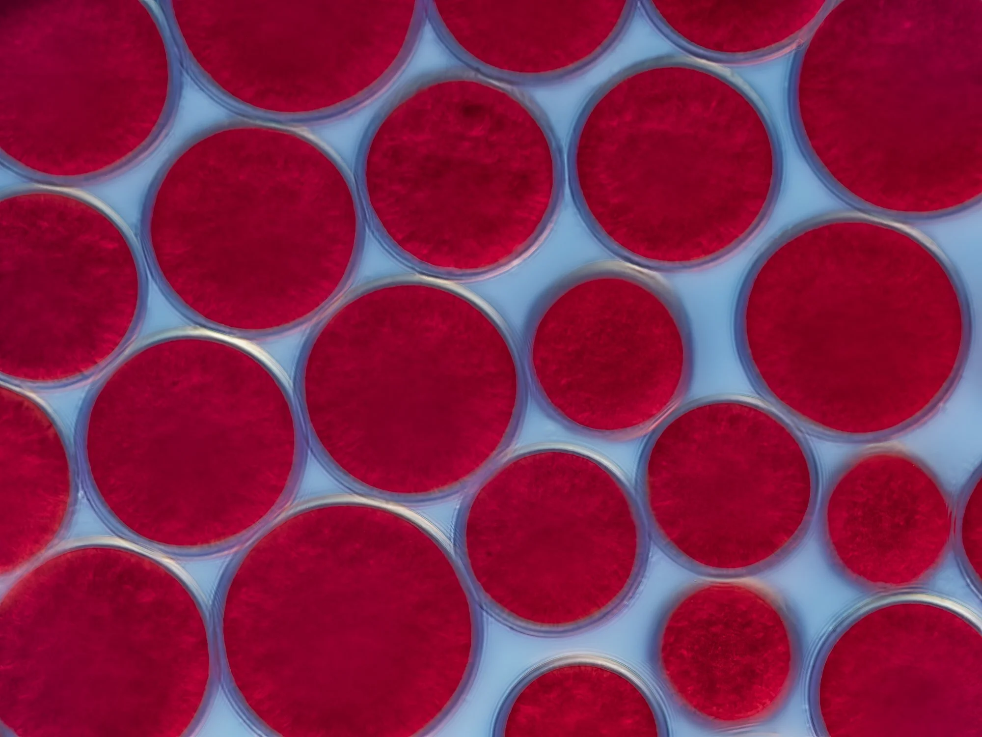
Light micrograph of red algae Polysiphonia sp. This photograph shows the top branches of the algae colony. Polysiphonia species are entirely marine, found growing on rock, other algae, mussels, etc. Red algae possess pigments of the phycoerythrine (red) and Phycocyanine (blue) group, involved in photosynthesis. Phycoerythrine reflects red light; hence the perception of the red color for us. The pigment absorbs rays in the blue-green-yellow range of the spectrum. These colors can penetrate deeper in the seawater than rays of other colors, and that is an advantage for the red algae. Magnification: x350 when printed at 10 centimetres wide at its longest edge.
Light micrograph of red algae Polysiphonia sp. This photograph shows the top branches of the algae colony. Polysiphonia species are entirely marine, found growing on rock, other algae, mussels, etc. Red algae possess pigments of the phycoerythrine (red) and Phycocyanine (blue) group, involved in photosynthesis. Phycoerythrine reflects red light; hence the perception of the red color for us. The pigment absorbs rays in the blue-green-yellow range of the spectrum. These colors can penetrate deeper in the seawater than rays of other colors, and that is an advantage for the red algae. Magnification: x325 when printed at 10 centimetres wide at its longest edge.
Aliquam erat volutpat. Quisque dapibus cursus magna vel mollis. Aliquam eu hendrerit nisl. Quisque vel accumsan urna. Nullam placerat sem non lectus eleifend mattis. Praesent gravida porta lectus, bibendum pharetra nibh efficitur vitae. Sed ut felis neque. Curabitur euismod diam ac neque rutrum, nec bibendum eros tempus. Ut aliquam aliquam lectus, nec sollicitudin urna hendrerit id. Quisque commodo in metus sit amet imperdiet. Cum sociis natoque penatibus et magnis dis parturient montes, nascetur ridiculus mus. Pellentesque id eros dignissim, porttitor ligula ac, rutrum leo. Integer ac vestibulum sapien, quis dignissim tellus. Sed quis cursus odio, placerat vehicula erat.
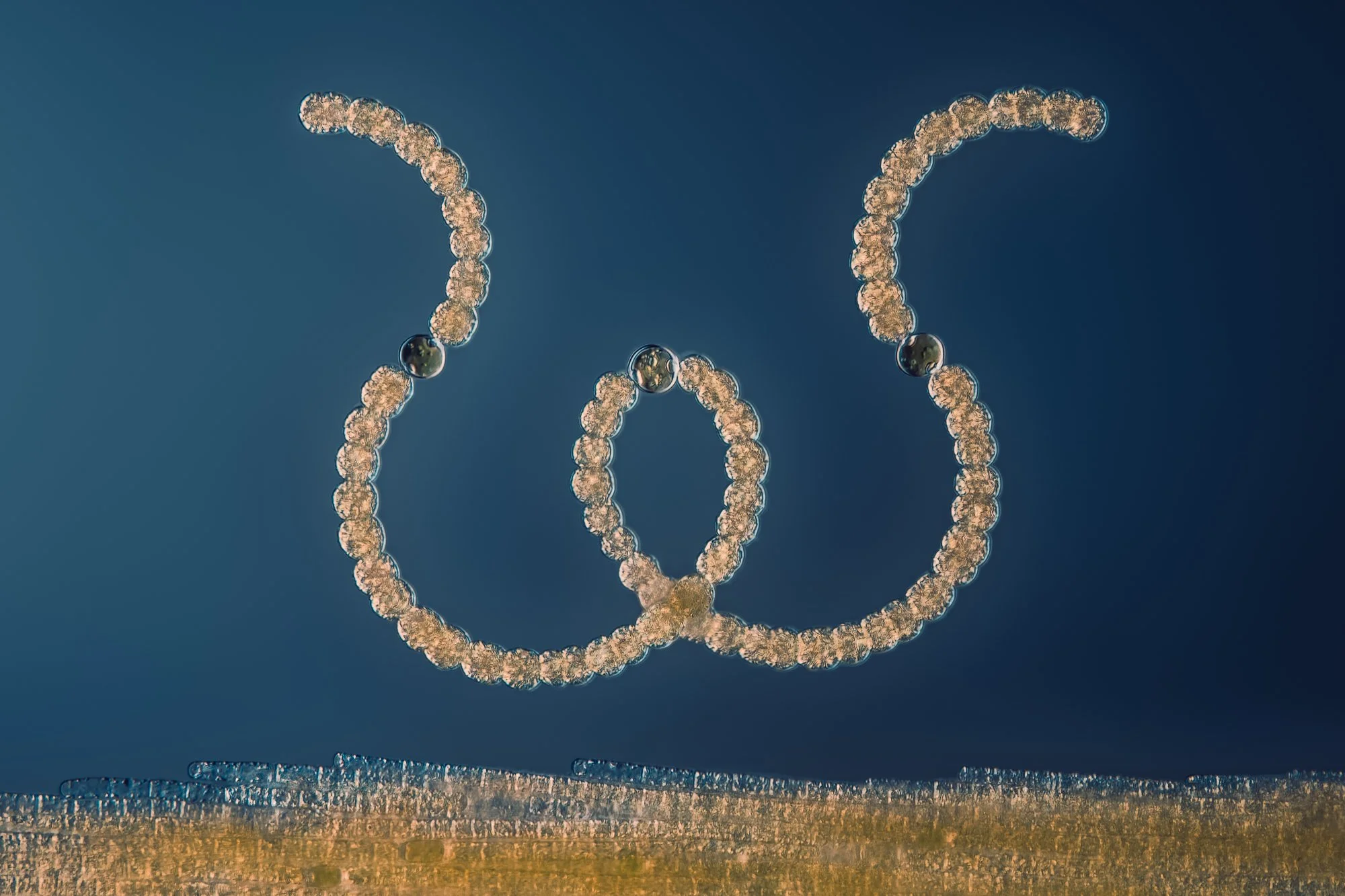
A light micrograph (fluorescent microscopy) of a colonial cyanobacteria Gloeotrichia sp. Gloeotrichia is a freshwater Cyanobacteria. In many parts of the world, Gloeotrichia appears unexpectedly in many lakes during late summer and fall as “Algae bloom". Gloeotrichia also has the ability to produce compounds called “microcystins” which are powerful toxins. There are many records of livestock, dogs and humans suffering liver damage through ingesting contaminated water. Magnification: x140 when printed at 10 centimetres wide at its longest edge.
A light micrograph (fluorescent microscopy) of a colonial cyanobacteria Gloeotrichia sp. Gloeotrichia is a freshwater Cyanobacteria. In many parts of the world, Gloeotrichia appears unexpectedly in many lakes during late summer and fall as “Algae bloom". Gloeotrichia also has the ability to produce compounds called “microcystins” which are powerful toxins. There are many records of livestock, dogs and humans suffering liver damage through ingesting contaminated water. Magnification: x275 when printed at 10 centimetres wide at its longest edge.
Magnification: x275 when printed at 10 centimetres wide at its longest edge.
A light micrograph (darkfield) of a colonial cyanobacteria Gloeotrichia sp. Gloeotrichia is a freshwater Cyanobacteria. In many parts of the world, Gloeotrichia appears unexpectedly in many lakes during late summer and fall as “Algae bloom". Gloeotrichia also has the ability to produce compounds called “microcystins” which are powerful toxins. There are many records of livestock, dogs and humans suffering liver damage through ingesting contaminated water. Magnification: x30 when printed at 10 centimetres wide at its longest edge.
Differential interference contrast (DIC) light micrograph of Chrococcus sp. Chroococcus sp. (color berry) is a cyanobacteria that is widely distributed in freshwater, less in saline waters. It is found in damp places such as moist soil, tree trunks, moist walls and rocks. The cells are single or united into spherical colonies, each containing a small number of cells surrounded by mucilage. Magnification: x425 when printed at 10 centimetres wide at its longest edge.
Differential interference contrast (DIC) light micrograph of Chrococcus sp. Chroococcus sp. (color berry) is a cyanobacteria that is widely distributed in freshwater, less in saline waters. It is found in damp places such as moist soil, tree trunks, moist walls and rocks. The cells are single or united into spherical colonies, each containing a small number of cells surrounded by mucilage. Magnification: x850 when printed at 10 centimetres wide at its longest edge.
Differential interference contrast (DIC) light micrograph of Spirulina sp. cyanobacteria filaments. Each filament is a colony of bacterial cells. Cyanobacteria are photosynthesizing bacteria that are found in most habitats where water is present. They are also used as a health food and dietary supplement because of their high protein content. Magnification: x550 when printed at 10 centimetres wide at its longest edge.
Light micrograph (fluorescence microscopy) of Spirulina sp. cyanobacteria filaments. Each filament is a colony of bacterial cells. Cyanobacteria are photosynthesizing bacteria that are found in most habitats where water is present. They are also used as a health food and dietary supplement because of their high protein content. Magnification: x400 when printed at 10 centimetres wide at its longest edge.
Light micrograph (darkfield) of a colony of cyanobacteria Aphanizomenon flos-aquae. It is one of the most common species of algae that is commonly used as a dietary supplement as it is a blue-green algae that can be commercially processed. It is known to contain various nutrients such as vitamins, amino acids, proteins, and phytochemicals. Magnification: x140 when printed at 10 centimetres wide at its longest edge.
Differential interference contrast light micrograph of cyanobacteria Dolichospermum sp. Dolichospermum sp. is a filamentous and nitrogen-fixing cyanobacterial genus that typically forms massive blooms in freshwater environments. Dolichospermum are capable of producing toxins. Magnification: x450 when printed at 10 centimetres wide at its longest edge.
Differential interference contrast (DIC) light micrograph of a cyanobacteria Woronichinia naegeliana. Woronichinia naegeliana can be found in freshwater nutrient-rich lakes and can form dense blooms. It is one of the most common species associated with toxic cyanobacteria bloom (algal blooms). Woronichinia naegeliana can produce anatoxins (nerve toxin), microcystins (liver toxin), microginins (liver toxin), and other less common toxins. There are gas vesicles in Woronichinia cells that provide a mechanism to move up and down in the water column, which increases access to nutrients and other growth factors. Magnification: x180 when printed at 10 centimetres wide at its longest edge.
Differential interference contrast micrograph of cyanobacteria Microcystis sp. Surrounding the colony there are rod-shaped bacteria that live in symbiosis with the Microcystis. The Microcystis is covered in a gel-like substance, mucilage, that provide a thriving habitat for the bacteria. The relationship between the bacteria and Microcystis is mutually beneficial. The associated bacterial flora depends on both carbon and energy sourced from the Microcystis, and supply the Microcystis with vitamin B12, which is required for the growth of the colony. The interactions between bacteria and cyanobacteria (blue-green algae) may play a significant part in the formation and development of algal blooms. The occurrence of cyanobacterial mass populations can create a significant water quality problem, especially as many cyanobacterial species are capable of synthesizing a wide range of odours, noxious compounds or potent toxins. Magnification: x450 when printed at 10 centimetres wide at its longest edge.
Differential interference contrast (DIC) light micrograph of a large terrestrial cyanobacteria (Stigonema mammilosum) often found growing on rocks. Stigonema mammilosum can be found as blackish crusts and biofilms on inselbergs (isolated rock hill, knob or ridge) in many places on earth. Stigonema mammilosum is a filamentous cyanobacteria with true branching (similar to plants) which gives it its characteristic appearance. Magnification: x425 when printed at 10 centimetres wide at its longest edge. when printed at 10 centimetres wide at its longest edge.
Differential interference contrast (DIC) light micrograph of a large terrestrial cyanobacteria (Stigonema mammilosum) often found growing on rocks. Stigonema mammilosum can be found as blackish crusts and biofilms on inselbergs (isolated rock hill, knob or ridge) in many places on earth. Stigonema mammilosum is a filamentous cyanobacteria with true branching (similar to plants) which gives it its characteristic appearance. Magnification: x350 when printed at 10 centimetres wide at its longest edge.
Differential interference contrast (DIC) light micrograph of a large terrestrial cyanobacteria (Stigonema mammilosum) often found growing on rocks. Stigonema mammilosum can be found as blackish crusts and biofilms on inselbergs (isolated rock hill, knob or ridge) in many places on earth. Stigonema mammilosum is a filamentous cyanobacteria with true branching (similar to plants) which gives it its characteristic appearance. Magnification: x425 when printed at 10 centimetres wide at its longest edge.
Differential interference contrast (DIC) light micrograph of Anabaena sp. algae (shape of W) and Aphanizomenon flos-aquae (bottom part) . Anabaena is a genus of filamentous cyanobacteria and are known for nitrogen-fixing abilities. Anabaena is capable to differentiate specialized cells, the heterocysts (round green cells in the photograph) to survive under different stress conditions. Under nitrogen limited condition, heterocysts provide the filament with nitrogen by fixing nitrogen. Both Anabaena and Aphanizomenon live in most freshwater lakes an ponds as well as in brackish water environments around the world. Magnification: x325 when printed at 10 centimetres wide at its longest edge.
Light micrograph (polarized light) of Anabaena sp. algae. Anabaena is a genus of filamentous cyanobacteria and are known for nitrogen-fixing abilities. They are one of four genera of cyanobacteria that produce neurotoxins, which are harmful to local wildlife, as well as farm animals and pets. Anabaena is capable to differentiate specialized cells, the heterocysts (round red/green cell in the photograph) to survive under different stress conditions. Under nitrogen limited condition, heterocysts provide the filament with nitrogen by fixing nitrogen. Magnification: x275 when printed at 10 centimetres wide at its longest edge.
Differential interference contrast (DIC) light micrograph of Anabaena sp. algae. Anabaena is a genus of filamentous cyanobacteria and are known for nitrogen-fixing abilities. They are one of four genera of cyanobacteria that produce neurotoxins, which are harmful to local wildlife, as well as farm animals and pets. Anabaena is capable to differentiate specialized cells, the heterocysts (round dark green cells) and akinetes (oval large cell), to survive under different stress conditions. Under nitrogen limited condition, heterocysts provide the filament with nitrogen by fixing nitrogen. Akinetes are spore-like dormant cells that allow survival during adverse environmental conditions. Magnification: x475 when printed at 10 centimetres wide at its longest edge.
Differential interference contrast (DIC) light micrograph of Merismopedia sp. Merismopedia is a genus of cyanobacteria found in fresh and salt water. It is ovoid or spherical in shape and arranged in rows and flats, forming rectangular colonies held together by a mucilaginous matrix. Species in this genus divide in only two directions, creating a characteristic grid-like pattern. Magnification: x140 when printed at 10 centimetres wide at its longest edge.
Differential interference contrast (DIC) light micrograph of Chrococcus sp. Chroococcus sp. (color berry) is a cyanobacteria that is widely distributed in freshwater, less in saline waters. It is found in damp places such as moist soil, tree trunks, moist walls and rocks. The cells are single or united into spherical colonies, each containing a small number of cells surrounded by mucilage. Magnification: x650 when printed at 10 centimetres wide at its longest edge.
Integer non elementum lectus. Nam eu sagittis lacus. Vestibulum aliquet bibendum tempus. Vestibulum bibendum, tortor sed ullamcorper suscipit, libero leo bibendum enim, nec posuere dui tellus vitae lacus. Mauris eget lectus velit. Aliquam et sem malesuada ligula iaculis tempus ac eget nulla. Ut ex quam, fringilla sed dolor ac, condimentum tempus nisl.
Vestibulum fringilla vitae velit sit amet pulvinar. Mauris sit amet nunc luctus, egestas est sit amet, suscipit nunc. Sed placerat velit quis commodo eleifend. Duis pulvinar neque tempor ex auctor ultricies. Nam sagittis consequat imperdiet. Cras hendrerit, quam in congue bibendum, libero tellus tempus massa, ut lacinia metus augue a nulla.
Banana fly. Coloured scanning electron micrograph (SEM). This image focuses on the head of the species Drosophila melanogaster, a pivotal species in genetic and biological research. The photograph reveals the compound eyes, antennae, and mouthparts, structures that are fundamental to the fly's sensory and feeding behaviors. D. melanogaster's head features, especially the large, red compound eyes, are critical for studying neurobiology and genetics, providing insights into vision, olfaction, and neural circuitry. The simplicity and genetic tractability of this organism have made it an invaluable model for dissecting the genetic underpinnings of development, behavior, and disease. This close-up view underscores the complexity and functionality of structures that are microscopic in size, yet monumental in scientific research. Magnification: x63 when printed 10 centimetres wide.
Banana fly. Coloured scanning electron micrograph (SEM). This image focuses on the mouth parts of the species Drosophila melanogaster, a pivotal species in genetic and biological research. The photograph reveals the mouth parts structures. The structure includes specialised organs such as the labellum, maxillae, and proboscis, adapted for sponging up liquid nutrients. D. melanogaster's mouthparts are a subject of study for understanding feeding behavior, sensory reception, and ecological interactions. These features reflect the evolutionary adaptations that facilitate the fruit fly's diet of decaying fruit and microbial content. The detailed examination of such morphological features aids in the broader understanding of insect physiology and the genetic basis of anatomical diversity. Magnification: x170 when printed 10 centimetres wide.
Banana fly. Coloured scanning electron micrograph (SEM). This image focuses on the mouth parts of the species Drosophila melanogaster, a pivotal species in genetic and biological research. The photograph reveals a close-up of mouth parts structures. D. melanogaster's mouthparts are a subject of study for understanding feeding behavior, sensory reception, and ecological interactions. These features reflect the evolutionary adaptations that facilitate the fruit fly's diet of decaying fruit and microbial content. The detailed examination of such morphological features aids in the broader understanding of insect physiology and the genetic basis of anatomical diversity. Magnification: x1850 when printed 10 centimetres wide.
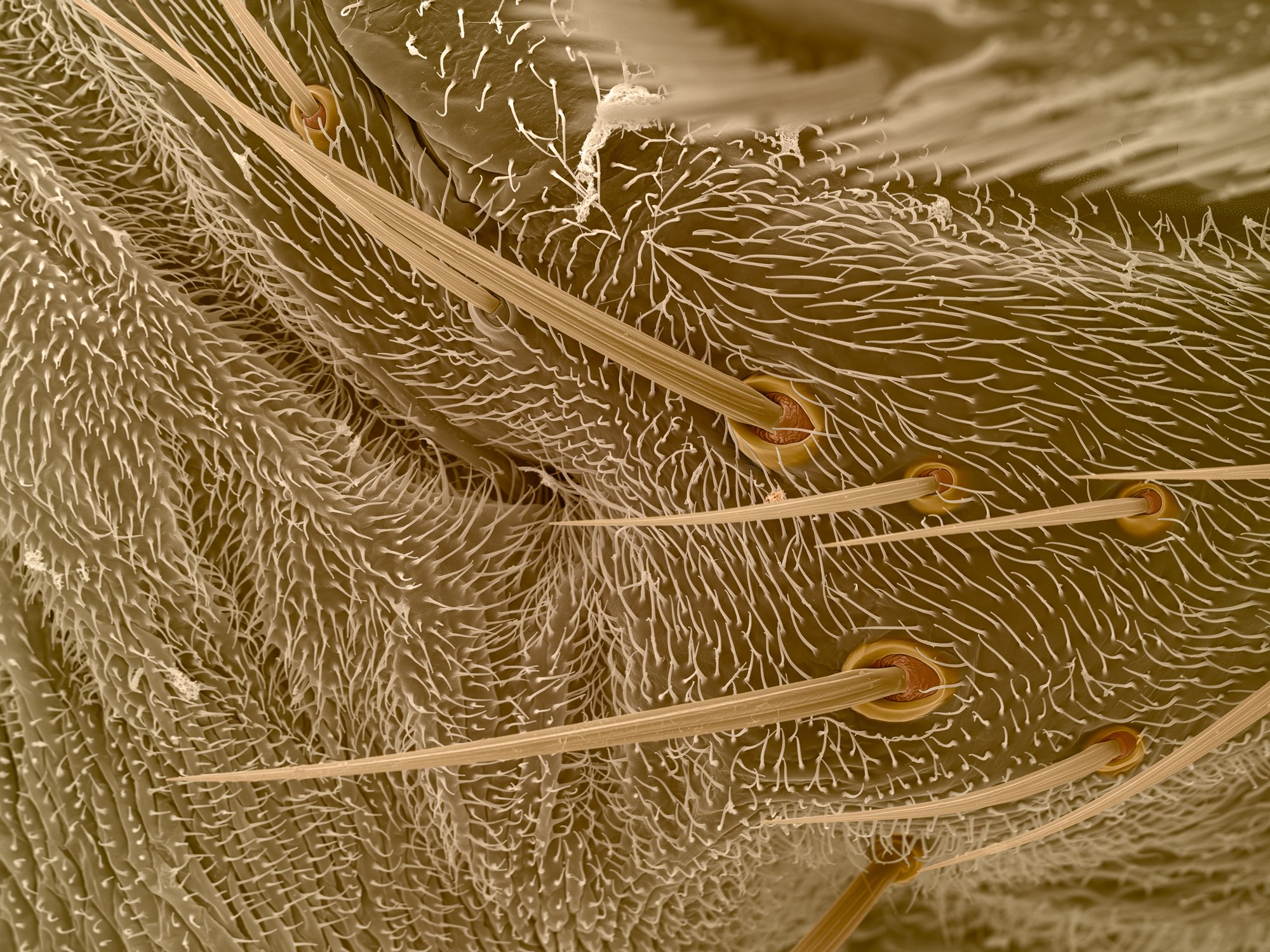
Banana fly. Coloured scanning electron micrograph (SEM). Detailed View of the species Drosophila melanogaster. This photograph provides a magnified look at the setae on the side of the head of a fruit flywith particular emphasis on the visible follicles from which the setae emerge. Setae, the hair-like sensory structures, play critical roles in the fly's ability to detect environmental cues, including chemical signals and physical touch. The image reveals the base of each seta, highlighting the follicle structure that anchors each seta to the exoskeleton of the fly. Such detailed visualization is essential for understanding the morphology and function of these sensory organs, contributing to our broader knowledge of insect sensory systems and their genetic and developmental underpinnings. Magnification: x450 when printed 10 centimetres
Light micrograph (darkfield) of a symbiotic relationship between microalgae and copepods. The microalgae are epibionts living on the surface of the copepod. Copepods are tiny crusteceans, about 1.5 mm in size. Normally, these species live separately, but in times when resources such as nutrition and oxygen levels are low, both benefit from a symbiotic relationship. Overgrown copepods, compared to those free from microalgae, are believed to have advantages in oxygen supply provided by the photosynthetic algae. At the same time, the copepod is the source of ammonium nitrogen the algae need. Magnification: x55 when printed at 10 centimetres wide at its longest edge.
Light micrograph (fluorescence microscopy) of a single water mite. Close-up. Water mites are tiny aquatic arachnids found in both freshwater and marine environments around the world. Magnification: x30 when printed at 10 centimetres wide at its longest edge.
Light micrograph (fluorescence microscopy) of a micromollusc. Micromolluscs are shelled mollusk which is extremely small even as adults. Many are only about 2 to 3 mm and a few have shells whichare as small as one millimeter. The photograph was captured using ultraviolet light. Microalgae and cyanobacteria that grow on the surface of the shell are autofluorescing in red and orange under the ultraviolet light. Magnification: x70 when printed at 10 centimetres wide at its longest edge.
Light micrograph (fluorescence microscopy) of a marine copepod (Tigriopus sp.). Copepods are the most abundant metazoan in the earth’s waters and are found in marine and fresh water, from lakes and streams to ocean trenches. They range in size from 0.1 to 10 mm and are a crucial link in the aquatic food web for fish and other planktivores. Magnification: x40 when printed at 10 centimetres wide at its longest edge.
Light micrograph (DIC) of a Rotifer Lepadella sp. Rotifers are the smallest animals on earth. Rotifers have existed for at least 35 million years and they only live for about 1-2 weeks. Under tough conditions (e.g. drought), rotifers can go into a desiccated state as a cyst or egg that can survive for decades without any signs of aging. The cysts can be carried by the wind and allow the rotifer to spread and escape harmful conditions. Once it finds water, the rotifers come back to life. Magnification: x425 when printed at 10 centimetres wide at its longest edge.
Light micrograph (polarized light) of Simocephalus sp. water flea. Water fleas are small crustaceans, commonly found in fresh water. They are filter feeders that ingest algae, protozoa or organic matter, and are a constituent of plankton. Magnification: x45 when printed at 10 centimetres wide at its longest edge.
Light micrograph (DIC) of a Rotifer Polyartha sp. Rotifers are the smallest animals on earth. The photograph shows a female with 5 nuclei in the vitallarium (a modified part of the ovary) and a yellow yolk. Rotifers have existed for at least 35 million years and they only live for about 1-2 weeks. Under tough conditions (e.g. drought), rotifers can go into a desiccated state as a cyst or egg that can survive for decades without any signs of aging. The cysts can be carried by the wind and allow the rotifer to spread and escape harmful conditions. Once it finds water, the rotifers come back to life. Magnification: x400 when printed at 10 centimetres wide at its longest edge.
Light micrograph (fluorescence microscopy) of a marine copepod (Tigriopus sp.). Copepods are the most abundant metazoan in the earth’s waters and are found in marine and fresh water, from lakes and streams to ocean trenches. They range in size from 0.1 to 10 mm and are a crucial link in the aquatic food web for fish and other planktivores. Magnification: x40 when printed at 10 centimetres wide at its longest edge.
Light micrograph (darkfield) of a rotifer Kellicottia longispina. Kellicottia longispina are rotifer species originating from North America and have become invasive in several continents. Magnification: x140 when printed at 10 centimetres wide at its longest edge.
Differential interference contrast (DIC) light micrograph nauplius larvae of a copepod. Copepods are the most abundant metazoan in the earth’s waters and are found in marine and fresh water, from lakes and streams to ocean trenches. They are a crucial link in the aquatic food web for fish and other planktivores. The larval stage have 3 pairs of legs and the typical red cyclops eye in the anterior centre of the animal. Magnification: x100 when printed at 10 centimetres wide at its longest edge.
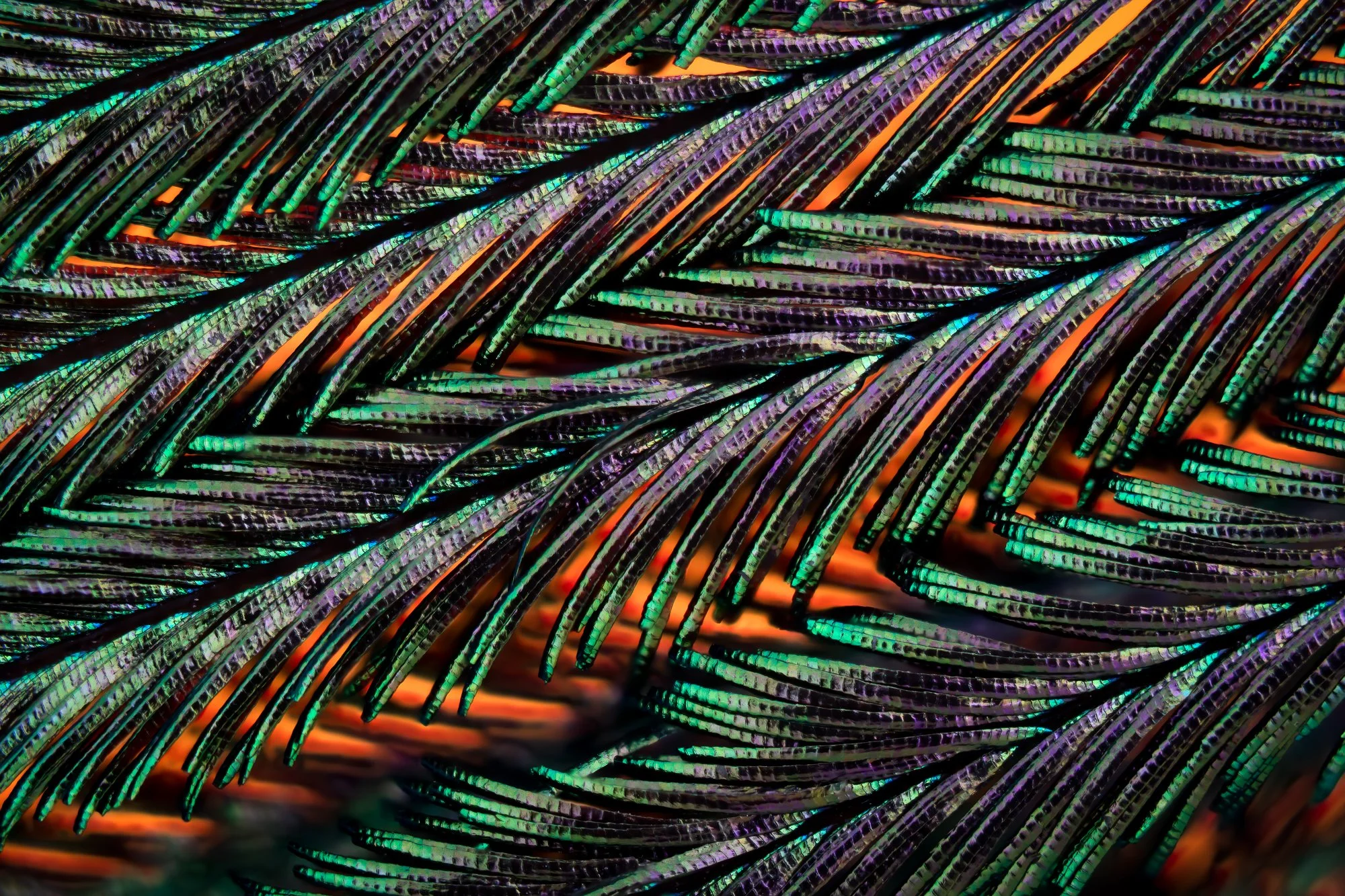
Light micrograph of a peacock plume feather. Peacocks achieve its stunning plumage display through structural coloration, more commonly known as iridescence. Structural coloration results from a lightwave interaction with the surface. Magnification: x30 when printed at 10 centimetres wide at its longest edge.
A Differential interference contrast (DIC) light micrgraph of a Rotifer of the genus Bdelloid. Also called wheel animals, rotifers are microscopic aquatic animals and common in freshwater environments. They are surprisingly complex on the inside: they have a brain, reproductive organs, a bladder and a digestive system. They are omnivorous, eating bacteria, debris or small protozoans. There are no male bdelloid rotifers. Females reproduce via parthenogenesis, a type of asexual reproduction. Magnification: x350 when printed at 10 centimetres wide at its longest edge.
Light micrograph of a peacock plume feather. Peacocks achieve its stunning plumage display through structural coloration, more commonly known as iridescence. Structural coloration results from a lightwave interaction with the surface. Magnification: x30 when printed at 10 centimetres wide at its longest edge.
Light micrograph of a peacock plume feather. Peacocks achieve its stunning plumage display through structural coloration, more commonly known as iridescence. Structural coloration results from a lightwave interaction with the surface. Magnification: x30 when printed at 10 centimetres wide at its longest edge.
Light micrograph (darkfield) of a Rotifer Keratella quadrata. Rotifers are among the smallest animals on earth. Rotifers have existed for at least 35 million years and they only live for about 1-2 weeks. Magnification: x110 when printed at 10 centimetres wide at its longest edge.
Sponge spicule. coloured scanning electron micrograph (SEM). A silica-based spicule from a sponge, a primitive aquatic organism that filters nutrients from water. Sponges, considered one of the most ancient and simple forms of life in aquatic environments, employ a filtration system to gather nutrients from the surrounding water. Their bodies are an assemblage of loosely organized cells, which gain structural integrity from spicules and fibers made predominantly of silica, and in some cases, calcium carbonate. The spicule depicted here exhibits a euaster form, characterized by its radial symmetry where extensions emanate from a central hub. Spicules play a crucial role in the sponge’s structural support and defense mechanisms against predators. Magnification: x2300 when printed 10 centimetres wide.
Light micrograph (DIC) of a Rotifer Brachionus sp. Rotifers are the smallest animals on earth. The photograph shows a female with an egg. Rotifers have existed for at least 35 million years and they only live for about 1-2 weeks. Magnification: x300 when printed at 10 centimetres wide at its longest edge.
Diffential interference contrast (DIC) light micrograph of Caenorhabditis elegans, a soil-dwelling bisexual nematode worm which feeds on bacteria. A tendency to reproduce by self-fertilization (resulting in identical offspring), along with the short time taken to reach maturity, make this tiny worm an ideal subject for genetic research. Scientists have already drawn up a 'wiring diagram' of its nervous system, studied the development of each of its 959 component cells and produced a map of its genes. Attempts are now being made to decode the genetic blueprint of C. elegans, to identify every one of the 100 million letters, or bases, in its genome. Magnification: x325 when printed at 10 centimetres wide at its longest edge.
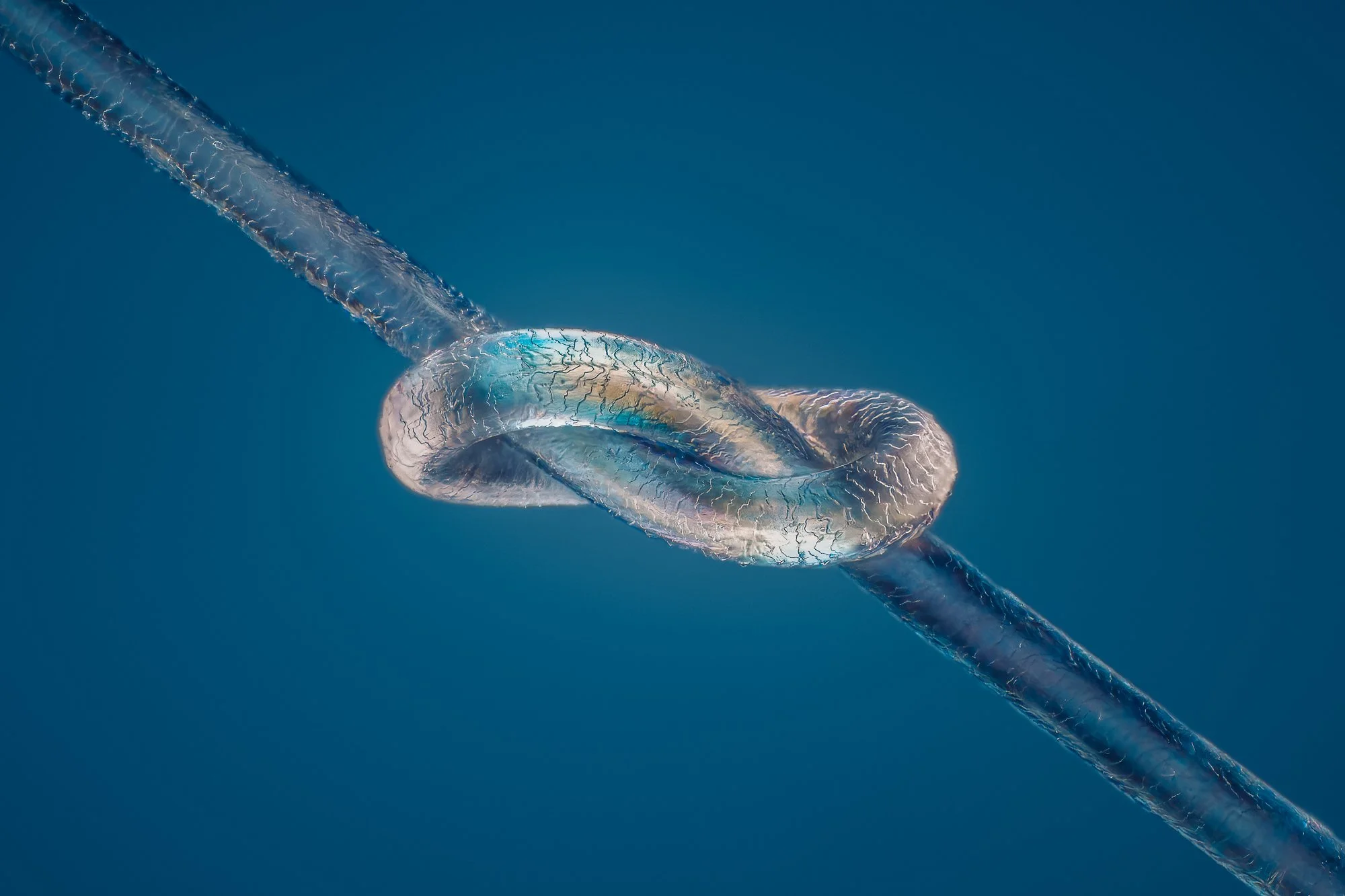
Light micrograph (polarized light) of a strand of human hair tied in a knot. Magnification: x325 when printed at 10 centimetres wide at its longest edge.
Light micrograph (polarized light) of a strand of human hair tied in a knot. Magnification: x190 when printed at 10 centimetres wide at its longest edge.
Light micrograph (fluorescent microscopy) of a Simocephalus sp. water flea's embryos. Water fleas are small crustaceans, commonly found in fresh water. They are filter feeders that ingest algae, protozoa or organic matter, and are a constituent of plankton. small crustaceans, commonly found in fresh water. They are filter feeders that ingest algae, protozoa or organic matter, and are a constituent of plankton. The larger black spots are the embryo's eyes. Magnification: x70 when printed at 10 centimetres wide at its longest edge.
Differential interference contrast (DIC) light micrograph of a strand of human hair. Magnification: x475 when printed at 10 centimetres wide at its longest edge.
Light micrograph of a Hydra. Hydras are small fresh-water animals with a tubular body. They are invertebrates where the free end of the body is a mouth opening surrounded by one to twelve thin, mobile tentacles. Each tentacle, or cnida is clothed with highly specialised stinging cells called cnidocytes. Cnidocytes contain specialized structures called nematocysts, which look like miniature light bulbs with a coiled thread inside. Upon contact with prey, the contents of the nematocyst are explosively discharged, firing a dart-like thread containing neurotoxins into whatever triggered the release which can paralyse the prey, especially if many hundreds of nematocysts are fired. Magnification: x35 when printed at 10 centimetres wide at its longest edge.
Light micrograph of a Hydra. Close-up of a tentacle. Hydras are small fresh-water animals with a tubular body. They are invertebrates where the free end of the body is a mouth opening surrounded by one to twelve thin, mobile tentacles. Each tentacle, or cnida is clothed with highly specialised stinging cells called cnidocytes. Cnidocytes contain specialized structures called nematocysts, which look like miniature light bulbs with a coiled thread inside. Upon contact with prey, the contents of the nematocyst are explosively discharged, firing a dart-like thread containing neurotoxins into whatever triggered the release which can paralyse the prey, especially if many hundreds of nematocysts are fired. Magnification: x275 when printed at 10 centimetres wide at its longest edge.
Light micrograph of a Hydra. Close-up of a tentacle with visible cells of nematocysts. Hydras are small fresh-water animals with a tubular body. They are invertebrates where the free end of the body is a mouth opening surrounded by one to twelve thin, mobile tentacles. Each tentacle, or cnida is clothed with highly specialised stinging cells called cnidocytes. Cnidocytes contain specialized structures called nematocysts, which look like miniature light bulbs with a coiled thread inside. Upon contact with prey, the contents of the nematocyst are explosively discharged, firing a dart-like thread containing neurotoxins into whatever triggered the release which can paralyse the prey, especially if many hundreds of nematocysts are fired. Magnification: x425 when printed at 10 centimetres wide at its longest edge.
Light micrograph of a Hydra. Close-up of a tentacle with visible cells of nematocysts. Hydras are small fresh-water animals with a tubular body. They are invertebrates where the free end of the body is a mouth opening surrounded by one to twelve thin, mobile tentacles. Each tentacle, or cnida is clothed with highly specialised stinging cells called cnidocytes. Cnidocytes contain specialized structures called nematocysts, which look like miniature light bulbs with a coiled thread inside. Upon contact with prey, the contents of the nematocyst are explosively discharged, firing a dart-like thread containing neurotoxins into whatever triggered the release which can paralyse the prey, especially if many hundreds of nematocysts are fired. Magnification: x1250 when printed at 10 centimetres wide at its longest edge.
Light micrograph (Composite image of fluorescent microscopy combined with brightfield) of a Simocephalus sp. water flea. Water fleas are small crustaceans, commonly found in fresh water. They are filter feeders that ingest algae, protozoa or organic matter, and are a constituent of plankton. The orange part in the middle are autofluorescing algae in the digestive system of the water flea. This water flea also carry embryos with visible eye-spots under her upper shell. embryos with visible eye-spots under her upper shell. Magnification: x20 when printed at 10 centimetres wide at its longest edge.
Light micrograph (darkfield with polarized light) of Simocephalus sp. water flea. Water fleas are small crustaceans, commonly found in fresh water. They are filter feeders that ingest algae, protozoa or organic matter, and are a constituent of plankton. light micrograph of Simocephalus sp. water flea. Water fleas are small crustaceans, commonly found in fresh water. They are filter feeders that ingest algae, protozoa or organic matter, and are a constituent of plankton. light micrograph of Simocephalus sp. water flea. Water fleas are small crustaceans, commonly found in fresh water. They are filter feeders that ingest algae, protozoa or organic matter, and are a constituent of plankton. Magnification: x45 when printed at 10 centimetres wide at its longest edge.
Light micrograph (polarized light microscopy) of a marine copepod (Tigriopus sp.). Copepods are the most abundant metazoan in the earth’s waters and are found in marine and fresh water, from lakes and streams to ocean trenches. They range in size from 0.1 to 10 mm and are a crucial link in the aquatic food web for fish and other planktivores. Magnification: x30 when printed at 10 centimetres wide at its longest edge.
Darkfield illuminated polarised light micrograph of a Cyclops sp. copepod carrying eggs (small, round). Copepods are microscopic aquatic crustaceans found in waters around the world. They may either be free living or parasitic on other organisms and are the main constituent of zooplankton. They feed on other microscopic animals and plants. Cyclops copepods are one of the most common freshwater copepods. Magnification: x70 when printed at 10 centimetres wide at its longest edge.
A light micrograph of an egg from a Brine shrimp (Artemia salina). This species is commonly found in saline waters such as salty lakes and salt pans. Magnification: x140 when printed at 10 centimetres wide at its longest edge.
Cras nec arcu ut nibh tincidunt venenatis. Suspendisse iaculis volutpat ultricies. Praesent congue metus ac nibh luctus feugiat. Quisque ac finibus velit. Fusce consectetur metus sed mauris consequat convallis. Pellentesque viverra eget mauris quis aliquam. Fusce lacinia auctor magna, vel vehicula sapien. Suspendisse eget augue ipsum. Aliquam luctus libero sed lacus iaculis volutpat. Pellentesque ullamcorper dolor a tincidunt interdum. Pellentesque ut commodo erat, sed fermentum nulla. Etiam sagittis congue hendrerit. Proin gravida vel purus quis efficitur. Ut faucibus vitae turpis quis consectetur.
Quisque et bibendum tellus. In hac habitasse platea dictumst. Sed vehicula egestas tortor, non luctus mi molestie eget. Vivamus at dolor non risus placerat pretium. Integer vitae cursus odio. Nulla facilisi. Suspendisse efficitur neque sit amet dui aliquam, ut ullamcorper mi accumsan. Integer egestas blandit rutrum. Morbi vel risus lectus. Quisque ultricies bibendum augue, sollicitudin auctor est maximus vitae. Nunc vitae ligula posuere, fringilla metus quis, facilisis nisl.
Ut id nunc vulputate, aliquet risus consectetur, cursus erat. Ut et porttitor diam. Nulla turpis massa, maximus id enim sed, aliquam porttitor neque. Praesent sit amet nibh sed magna dapibus finibus. In et sollicitudin mi, eget vulputate ligula. In rutrum ultrices ipsum a bibendum. Morbi luctus quis odio eget fringilla.
Mauris rhoncus ex elit, ac finibus velit dignissim et. Vivamus imperdiet pharetra facilisis. Donec blandit aliquam libero, vel euismod massa ultricies id. Vestibulum venenatis dignissim venenatis. Nullam sagittis faucibus pellentesque. Duis suscipit enim a tellus convallis ultricies. Donec vehicula eleifend dictum. Nam vel auctor leo, a bibendum massa. Phasellus nulla turpis, auctor eu vulputate at.
Differential interference contrast (DIC) light micrograph of Paramecium sp. Paramecium is a ciliate protozoan, with oral groove, food vacuoles, nucleus, and cilia. Paramecium are found mainly in stagnant ponds, feeding on bacteria and plant particles. They have a permanent mouth called an oral grove. Food taken in through the oral groove is digested within temporary digestive vacuoles in the cell cytoplasm. The two contractile vacuoles act as osmotic regulators, controlling the flow of water across the ciliate's membrane. The contractile vacuoles are surrounded by a halo of radiating channels. Excess water collects in the channels, drains into the vacuole and is then discharged through a pore at the surface. Magnification: x170 when printed at 10 centimetres wide at its longest edge.
Differential interference contrast (DIC) light micrograph of Nassula sp. Nassula is a genus of unicellular ciliates that belongs to the Nassophorea class. Like other members of this group, it has a basket-like feeding apparatus (cytopharynx) that consists of a series of microtubules (seen in the photograph). These are designed to allow the organism to ingest and transport nutrients from filamentous blue-green algae. The organisms use this structure to draw individual strands of algae through the cytopharynx and into the body. Magnification: x325 when printed at 10 centimetres wide at its longest edge.
Amoeba (Arcella). Like a book-illustration with all internal features. Nuclei, pseudopoda, episode, vacuoles and food. Also, the shell is visible.
Differential interference contrast light micrograph of a Vorticella sp. ciliate protozoa. Vorticella are mostly sessile, meaning they attach to a surface and cannot move around on their own. When disturbed, they contract their stalk into a tight coil. This contraction is one of the fastest known actions of any species. Magnification: x275 when printed at 10 centimetres wide at its longest edge.
Differential interference contrast (DIC) light micrograph of Lepocinclis acus. micrograph shows the paramylons of Lepocinclis acus, which are tube-like structures that are used as power reserves. They are produced by the photosynthesis process. A red eye spot known as stigma helps the cell swim toward favorable conditions. Magnification: x425 when printed at 10 centimetres wide at its longest edge.
Differential interference contrast (DIC) light micrograph of a Stentor sp. freshwater ciliate protozoan. The body, or cortex, is generally trumpet-shaped with a ring of prominent cilia that sweep in food and aid in swimming. Some Stentor species can grow up to two millimetres in length, making them among the largest single-celled organisms. Magnification: x95 when printed at 10 centimetres wide at its longest edge.
Differential interference contrast light micrograph of a Vorticella sp. ciliate protozoa. Vorticella are mostly sessile, meaning they attach to a surface and cannot move around on their own. When disturbed, they contract their stalk into a tight coil. This contraction is one of the fastest known actions of any species. Magnification: x575 when printed at 10 centimetres wide at its longest edge.
Cras tortor augue, dapibus vel viverra at, mattis sit amet lectus. Aliquam odio nibh, rhoncus a lectus vitae, finibus venenatis massa. Nullam mattis, nisi quis tempus interdum, metus neque malesuada enim, eget ullamcorper magna tortor at massa. Fusce feugiat scelerisque dui, nec faucibus ligula efficitur faucibus.
Phasellus pretium consectetur pharetra. Maecenas vel rutrum nisi. Sed hendrerit, diam sed eleifend interdum, felis nisl aliquam enim, vestibulum feugiat lectus eros ut leo. Maecenas aliquet tortor nec lacus viverra convallis. Praesent dignissim nulla id dolor mollis, ut suscipit neque sollicitudin. Suspendisse lacinia, mi a venenatis porta, magna nulla consectetur eros, vel ultricies arcu elit ornare est. Donec eu mi nunc. Vivamus diam eros, molestie nec rutrum ut, tempus eget turpis.
Reindeer lichen. Coloured scanning electron micrograph (SEM) of Reindeer lichen (Cladonia rangiferina). Magnified to reveal intricate details of its thallus structure. The image showcases the lichen's branching morphology and surface texture. It thrives in cold and arid environments. Reindeer lichen is characterised by its symbiotic relationship between the fungal and algal components, critical for the lichen's photosynthesis and nutrient uptake. Reindeer lichen is a vital food source for caribou and reindeer, plays a significant ecological role in soil formation and nutrient cycling, and is notable for its extremely slow growth rate, often less than 5 millimeter per year, making it vulnerable to overgrazing and environmental changes. Magnification: x50 when printed 10 centimetres wide.
Reindeer lichen. Coloured scanning electron micrograph (SEM) of Reindeer lichen (Cladonia rangiferina). Magnified to reveal intricate details of its thallus structure. The image showcases the lichen's branching morphology and surface texture. It thrives in cold and arid environments. Reindeer lichen is characterised by its symbiotic relationship between the fungal and algal components, critical for the lichen's photosynthesis and nutrient uptake. Reindeer lichen is a vital food source for caribou and reindeer, plays a significant ecological role in soil formation and nutrient cycling, and is notable for its extremely slow growth rate, often less than 5 millimeter per year, making it vulnerable to overgrazing and environmental changes. Magnification: x23 when printed 10 centimetres wide. when printed 10 centimetres wide.
Reindeer lichen. Coloured scanning electron micrograph (SEM) of Reindeer lichen (Cladonia rangiferina). Magnified to reveal intricate details of its thallus structure. The image showcases the lichen's branching morphology and surface texture. It thrives in cold and arid environments. Reindeer lichen is characterised by its symbiotic relationship between the fungal and algal components, critical for the lichen's photosynthesis and nutrient uptake. Reindeer lichen is a vital food source for caribou and reindeer, plays a significant ecological role in soil formation and nutrient cycling, and is notable for its extremely slow growth rate, often less than 5 millimeter per year, making it vulnerable to overgrazing and environmental changes. Magnification: x180 when printed 10 centimetres wide.
Reindeer lichen. Coloured scanning electron micrograph (SEM) of Reindeer lichen (Cladonia rangiferina). Magnified to reveal intricate details of its thallus structure. The image showcases the lichen's branching morphology and surface texture. It thrives in cold and arid environments. Reindeer lichen is characterised by its symbiotic relationship between the fungal and algal components, critical for the lichen's photosynthesis and nutrient uptake. Reindeer lichen is a vital food source for caribou and reindeer, plays a significant ecological role in soil formation and nutrient cycling, and is notable for its extremely slow growth rate, often less than 5 millimeter per year, making it vulnerable to overgrazing and environmental changes. Magnification: x155 when printed 10 centimetres wide.
Light micrograph (polarized light) of crystallized Sodium phenylbyturate (Ammonaps). Sodium phenylbutyrate is used to help treat urea cycle disorders. Magnification: x50 when printed at 10 centimetres wide at its longest edge.
Light micrograph (polarized light) of crystallized Sodium phenylbyturate (Ammonaps). Sodium phenylbutyrate is used to help treat urea cycle disorders. Magnification: x50 when printed at 10 centimetres wide at its longest edge.
Light micrograph (polarized light) of crystallized Sodium phenylbyturate (Ammonaps). Sodium phenylbutyrate is used to help treat urea cycle disorders.Magnification: x50 when printed at 10 centimetres wide at its longest edge.
Light micrograph (polarized light) of crystallized Sodium phenylbyturate (Ammonaps). Sodium phenylbutyrate is used to help treat urea cycle disorders. Magnification: x50 when printed at 10 centimetres wide at its longest edge.
Light micrograph (polarized light) of crystallized Sodium phenylbyturate (Ammonaps). Sodium phenylbutyrate is used to help treat urea cycle disorders. Magnification: x50 when printed at 10 centimetres wide at its longest edge.
Light micrograph (polarized light) of crystallized Sodium phenylbyturate (Ammonaps). Sodium phenylbutyrate is used to help treat urea cycle disorders. Magnification: x50 when printed at 10 centimetres wide at its longest edge.
Light micrograph (polarized light) of crystallized Sodium phenylbyturate (Ammonaps). Sodium phenylbutyrate is used to help treat urea cycle disorders. Magnification: x100 when printed at 10 centimetres wide at its longest edge.
Light micrograph (polarized light) of crystallized Sodium phenylbyturate (Ammonaps). Sodium phenylbutyrate is used to help treat urea cycle disorders. Magnification: x100 when printed at 10 centimetres wide at its longest edge.
Light micrograph (polarized light) of crystallized Sodium phenylbyturate (Ammonaps). Sodium phenylbutyrate is used to help treat urea cycle disorders. Magnification: x50 when printed at 10 centimetres wide at its longest edge.
Diatoms, coloured scanning electron micrograph (SEM). Coscinodiscus marginatus (left) and Eupyxidicula sp. (right). Shows to the intricate silica-based cell walls of two fossil diatoms, showcasing the beauty and complexity of these microscopic organisms. Diatoms, a major group of algae found in the oceans, waterways, and soils of the world, are not just marvels of natural art; they are critical to our planet's ecology. They contribute significantly to the oxygen in the atmosphere and are a fundamental link in the aquatic food chain. Diatoms are among the most diverse species on earth. Estimates of the number of diatom species range from 20.000-200.000. Scientists are discovering new species every year. Magnification: x600 when printed 10 centimetres wide.
Lily pollen. Coloured scanning electron micrograph (SEM). The distinctive texturing on each pollen grain is designed to enhance adherence to insects, such as bees and butterflies, that visit the flowers. These structural adaptations are critical for ensuring that pollen is efficiently transferred between flowers, aiding in cross-pollination. The allergenic properties of lily pollen are notable. Despite their microscopic size, these grains can have a profound impact on human health, triggering allergic reactions in susceptible individuals. Magnification: x470 when printed 10 centimetres wide.
Head louse (Pediculus humanus capitis). Coloured scanning electron micrograph. The head louse (Pediculus humanus capitis) is a small insect, usually 1-2 mm long, with a large abdomen and legs equipped with sharp claws for gripping hair. Lacking wings, these parasites have a slightly narrower head than their body and use their piercing mouthparts to dig into the skin, draining blood. Thriving on the human scalp for over 100,000 years, they cause itching and discomfort without transmitting diseases. Magnification: x190 when printed 10 centimetres wide.
Head louse (Pediculus humanus capitis). Coloured scanning electron micrograph. The head louse (Pediculus humanus capitis) is a small insect, usually 1-2 mm long, with a large abdomen and legs equipped with sharp claws for gripping hair. Lacking wings, these parasites have a slightly narrower head than their body and use their piercing mouthparts to dig into the skin, draining blood. Thriving on the human scalp for over 100,000 years, they cause itching and discomfort without transmitting diseases. Magnification: 440x when printed 10 centimetres wide.
Head louse (Pediculus humanus capitis). Coloured scanning electron micrograph. The head louse (Pediculus humanus capitis) is a small insect, usually 1-2 mm long, with a large abdomen and legs equipped with sharp claws for gripping hair. Lacking wings, these parasites have a slightly narrower head than their body and use their piercing mouthparts to dig into the skin, draining blood. Thriving on the human scalp for over 100,000 years, they cause itching and discomfort without transmitting diseases. Magnification: x80 when printed 10 centimetres wide.
Fruit fly. Coloured scanning electron micrograph (SEM). This image focuses on the head of the species Drosophila melanogaster, a pivotal species in genetic and biological research. The photograph reveals the compound eyes, antennae, and mouthparts, structures that are fundamental to the fly's sensory and feeding behaviors. D. melanogaster's head features, especially the large, red compound eyes, are critical for studying neurobiology and genetics, providing insights into vision, olfaction, and neural circuitry. The simplicity and genetic tractability of this organism have made it an invaluable model for dissecting the genetic underpinnings of development, behavior, and disease. This close-up view underscores the complexity and functionality of structures that are microscopic in size, yet monumental in scientific research. Magnification: x63 when printed 10 centimetres wide.
Fruit fly. Coloured scanning electron micrograph (SEM). This image focuses on the mouth parts of the species Drosophila melanogaster, a pivotal species in genetic and biological research. The photograph reveals the mouth parts structures. The structure includes specialised organs such as the labellum, maxillae, and proboscis, adapted for sponging up liquid nutrients. D. melanogaster's mouthparts are a subject of study for understanding feeding behavior, sensory reception, and ecological interactions. These features reflect the evolutionary adaptations that facilitate the fruit fly's diet of decaying fruit and microbial content. The detailed examination of such morphological features aids in the broader understanding of insect physiology and the genetic basis of anatomical diversity. Magnification: x170 when printed 10 centimetres wide.
Fruit fly. Coloured scanning electron micrograph (SEM). This image focuses on the mouth parts of the species Drosophila melanogaster, a pivotal species in genetic and biological research. The photograph reveals a close-up of mouth parts structures. D. melanogaster's mouthparts are a subject of study for understanding feeding behavior, sensory reception, and ecological interactions. These features reflect the evolutionary adaptations that facilitate the fruit fly's diet of decaying fruit and microbial content. The detailed examination of such morphological features aids in the broader understanding of insect physiology and the genetic basis of anatomical diversity. Magnification: x1850 when printed 10 centimetres wide.
Fruit fly. Coloured scanning electron micrograph (SEM). Detailed View of the species Drosophila melanogaster. This photograph provides a magnified look at the setae on the side of the head of a fruit flywith particular emphasis on the visible follicles from which the setae emerge. Setae, the hair-like sensory structures, play critical roles in the fly's ability to detect environmental cues, including chemical signals and physical touch. The image reveals the base of each seta, highlighting the follicle structure that anchors each seta to the exoskeleton of the fly. Such detailed visualization is essential for understanding the morphology and function of these sensory organs, contributing to our broader knowledge of insect sensory systems and their genetic and developmental underpinnings. Magnification: x450 when printed 10 centimetres
Fruit fly's eye. Coloured scanning electron micrograph (SEM). This image showcases the compound eye structure of Drosophila melanogaster, commonly known as the fruit fly. The micrograph reveals the highly organized array of ommatidia, the individual photoreceptive units that make up the compound eye. Each ommatidium consists of a cluster of cells that work together to capture and process light, contributing to the fly's ability to detect movement and form images.Magnification: x450 when printed 10 centimetres wide.
Diatom. Coloured scanning electron micrograph (SEM). The photograph shows the genus Gomphonema sp. Diatoms contribute significantly to the oxygen in the atmosphere and are a fundamental link in the aquatic food chain. Diatoms are among the most diverse species on earth. Estimates of the number of diatom species range from 20.000-200.000. Scientists are discovering new species every year. Magnification: x1260 when printed 10 centimetres wide
Sponge spicule. coloured scanning electron micrograph (SEM). A silica-based spicule from a sponge, a primitive aquatic organism that filters nutrients from water. Sponges, considered one of the most ancient and simple forms of life in aquatic environments, employ a filtration system to gather nutrients from the surrounding water. Their bodies are an assemblage of loosely organized cells, which gain structural integrity from spicules and fibers made predominantly of silica, and in some cases, calcium carbonate. The spicule depicted here exhibits a euaster form, characterized by its radial symmetry where extensions emanate from a central hub. Spicules play a crucial role in the sponge’s structural support and defense mechanisms against predators. Magnification: x2300 when printed 10 centimetres wide.
Diatom. Coloured scanning electron micrograph (SEM). Species Didymosphenia geminata, inside valve view. Also colloquially known as 'rock snot' due to its capability to form thick, mucilaginous mats on the bottom of rivers and streams. Characterized by its stalked colonies and distinctive morphology, D. geminata is a subject of ecological concern because of its tendency to bloom excessively under certain environmental conditions. These blooms can cover extensive areas of substrate, impacting aquatic ecosystems by altering habitat structures, nutrient cycling, and water flow. Diatoms contribute significantly to the oxygen in the atmosphere and are a fundamental link in the aquatic food chain. Diatoms are among the most diverse species on earth. Estimates of the number of diatom species range from 20.000-200.000. Scientists are discovering new species every year. Magnification: x680 when printed 10 centimetres wide.
Diatom. Coloured scanning electron micrograph (SEM). Species Didymosphenia geminata, Close-up of outside valve view. Also colloquially known as 'rock snot' due to its capability to form thick, mucilaginous mats on the bottom of rivers and streams. Characterized by its stalked colonies and distinctive morphology, D. geminata is a subject of ecological concern because of its tendency to bloom excessively under certain environmental conditions. These blooms can cover extensive areas of substrate, impacting aquatic ecosystems by altering habitat structures, nutrient cycling, and water flow. Diatoms contribute significantly to the oxygen in the atmosphere and are a fundamental link in the aquatic food chain. Diatoms are among the most diverse species on earth. Estimates of the number of diatom species range from 20.000-200.000. Scientists are discovering new species every year. Magnification: x5500 when printed 10 centimetres wide.
Diatom. Coloured scanning electron micrograph (SEM). The photograph shows the genus Gomphonema sp. Diatoms contribute significantly to the oxygen in the atmosphere and are a fundamental link in the aquatic food chain. Diatoms are among the most diverse species on earth. Estimates of the number of diatom species range from 20.000-200.000. Scientists are discovering new species every year. Magnification: x1260 when printed 10 centimetres wide
Reindeer lichen. Coloured scanning electron micrograph (SEM) of Reindeer lichen (Cladonia rangiferina). Magnified to reveal intricate details of its thallus structure. The image showcases the lichen's branching morphology and surface texture. It thrives in cold and arid environments. Reindeer lichen is characterised by its symbiotic relationship between the fungal and algal components, critical for the lichen's photosynthesis and nutrient uptake. Reindeer lichen is a vital food source for caribou and reindeer, plays a significant ecological role in soil formation and nutrient cycling, and is notable for its extremely slow growth rate, often less than 5 millimeter per year, making it vulnerable to overgrazing and environmental changes. Magnification: x23 when printed 10 centimetres wide. when printed 10 centimetres wide.
Reindeer lichen. Coloured scanning electron micrograph (SEM) of Reindeer lichen (Cladonia rangiferina). Magnified to reveal intricate details of its thallus structure. The image showcases the lichen's branching morphology and surface texture. It thrives in cold and arid environments. Reindeer lichen is characterised by its symbiotic relationship between the fungal and algal components, critical for the lichen's photosynthesis and nutrient uptake. Reindeer lichen is a vital food source for caribou and reindeer, plays a significant ecological role in soil formation and nutrient cycling, and is notable for its extremely slow growth rate, often less than 5 millimeter per year, making it vulnerable to overgrazing and environmental changes. Magnification: x23 when printed 10 centimetres wide.
Reindeer lichen. Coloured scanning electron micrograph (SEM) of Reindeer lichen (Cladonia rangiferina). Magnified to reveal intricate details of its thallus structure. The image showcases the lichen's branching morphology and surface texture. It thrives in cold and arid environments. Reindeer lichen is characterised by its symbiotic relationship between the fungal and algal components, critical for the lichen's photosynthesis and nutrient uptake. Reindeer lichen is a vital food source for caribou and reindeer, plays a significant ecological role in soil formation and nutrient cycling, and is notable for its extremely slow growth rate, often less than 5 millimeter per year, making it vulnerable to overgrazing and environmental changes. Magnification: x155 when printed 10 centimetres wide.
Reindeer lichen. Coloured scanning electron micrograph (SEM) of Reindeer lichen (Cladonia rangiferina). Magnified to reveal intricate details of its thallus structure. The image showcases the lichen's branching morphology and surface texture. It thrives in cold and arid environments. Reindeer lichen is characterised by its symbiotic relationship between the fungal and algal components, critical for the lichen's photosynthesis and nutrient uptake. Reindeer lichen is a vital food source for caribou and reindeer, plays a significant ecological role in soil formation and nutrient cycling, and is notable for its extremely slow growth rate, often less than 5 millimeter per year, making it vulnerable to overgrazing and environmental changes. Magnification: x180 when printed 10 centimetres wide.
Diatom (Neidium sp). Coloured scanning electron micrograph (SEM). Neidium sp. Diatoms are distinguished by their valve shapes, which vary from rounded to capitate, rostrate, or protracted ends, adapted across different species. Thriving predominantly in slightly acidic waters, Neidium, though not abundantly found, plays a pivotal role in aquatic ecosystems and is widely distributed. Diatoms contribute significantly to the oxygen in the atmosphere and are a fundamental link in the aquatic food chain. Diatoms are among the most diverse species on earth. Estimates of the number of diatom species range from 20.000-200.000. Scientists are discovering new species every year. Magnification: x1520 when printed 10 centimetres wide.
Fossil Diatom (Suriella sp.) surrounded by various cracked diatom and radiolaria parts. Coloured scanning electron micrograph (SEM). Diatoms contribute significantly to the oxygen in the atmosphere and are a fundamental link in the aquatic food chain. Diatoms are among the most diverse species on earth. Estimates of the number of diatom species range from 20.000-200.000. Scientists are discovering new species every year. Magnification: x870 when printed 10 centimetres wide.
Fossil Diatom (Thalassiosira sp.) surrounded by various cracked diatom and radiolaria parts. Coloured scanning electron micrograph (SEM). Diatoms contribute significantly to the oxygen in the atmosphere and are a fundamental link in the aquatic food chain. Diatoms are among the most diverse species on earth. Estimates of the number of diatom species range from 20.000-200.000. Scientists are discovering new species every year. Magnification: x170 when printed 10 centimetres wide.
SEM
Fossil Diatom (Biddulphia sp.) surrounded by various cracked diatom and radiolaria parts. Coloured scanning electron micrograph (SEM). Diatoms contribute significantly to the oxygen in the atmosphere and are a fundamental link in the aquatic food chain. Diatoms are among the most diverse species on earth. Estimates of the number of diatom species range from 20.000-200.000. Scientists are discovering new species every year. Magnification: x530 when printed 10 centimetres wide.
Fossil Diatom (Biddulphia sp.) surrounded by various cracked diatom and radiolaria parts. Coloured scanning electron micrograph (SEM). Diatoms contribute significantly to the oxygen in the atmosphere and are a fundamental link in the aquatic food chain. Diatoms are among the most diverse species on earth. Estimates of the number of diatom species range from 20.000-200.000. Scientists are discovering new species every year. Magnification: x530 when printed 10 centimetres wide.
Haematococcus pluvalis. DIC
Haematococcus pluvialis in its vibrant red stage, showcased in this close-up image. Known for transitioning from green to red under stress, this green algae accumulates astaxanthin, a powerful antioxidant. As a popular dietary supplement, it offers numerous health benefits, including enhanced skin health and immune support. The intense red color indicates the algae's high astaxanthin content, making it a valuable component in nutritional products.
Light micrograph (polarized light) of crystallized Oxycodone (dihydrohydroxycodeinone). Oxycodone is an opioid medication and commonly prescribed for acute pain conditions but is also known for its high potential for dependency and misuse, leading to significant public health concerns related to opioid addiction. Crystalized and photographed in polarized light with retarders. Magnification: x140 when printed 10 centimetres wide.
Amoeba (Arcella). Like a book-illustration with all internal features. Nuclei, pseudopoda, episode, vacuoles and food. Also, the shell is visible.